 |
|
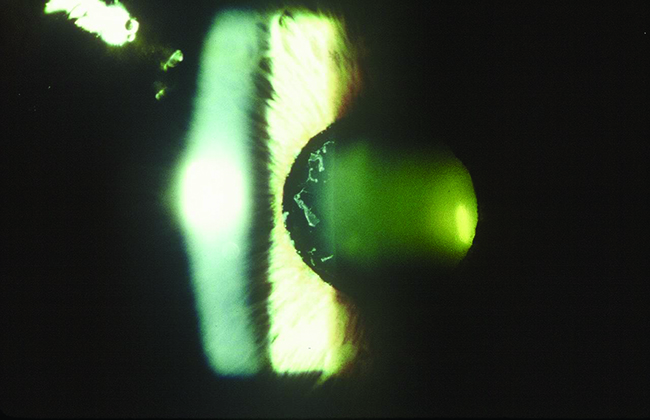
In non-glaucomatous XFS, the vascular supply defect plays a more critical role than intraocular pressure. However, with other forms of the pseudoexfoliation spectrum, intraocular pressure adds to risk factors of damage. Photo: Paul Ajamian, OD. Click image to enlarge. |
Unlike primary open-angle glaucoma, with its usually slowly progressive and fairly predictable course, the pseudoexfoliative form can be more aggressive and difficult to control. For this reason, it’s important to be vigilant for the presence of pseudoexfoliation syndrome (XFS) as a precursor to the development of secondary glaucoma. One recent study aimed to assess macular and retinal nerve fiber layer (RNFL) thickness, as well as the somewhat newer metric of Bruch’s membrane opening minimum rim width (BMO-MRW), in those with non-glaucomatous XFS were compared with controls to evaluate predictors of glaucomatous damage.
BMO-MRW is the shortest distance between the internal limiting membrane and interior opening of Bruch’s membrane; it has recently been considered a more accurate reflection of neuroretinal rim integrity and better predictor of visual field status than existing glaucoma indicators like RNFL thickness.
The prospective study used OCT to evaluate 32 eyes of 32 individuals with clinically detected non-glaucomatous XFS, along with 30 eyes of age- and sex-matched controls. Results indicated that no difference was seen between the cases and controls in intraocular pressure, with measurements on average of 14.9mm Hg in patients and 15.3mm Hg in controls. However, the BMO-MRW of the superotemporal segment was found to be thinner in the XFS group than in controls, on average with measurements of 303.7 ±60.5µm and 341.4 ±56.2µm, respectively. This was the only main difference observed, as no other discrepancies were seen in the other MRW sectors, inner macular layers and circumpapillary RNFL thickness between groups.
In a recent paper on the work for Journal of Glaucoma, the researchers discussed further how these results might be indicative of early glaucoma in these patients. As they explain, glaucomatous damage can arise in XFS thought elevated intraocular pressure, diminishment of blood flow, and hemodynamic changes in the retina, optic nerve head and choroidal and retrobulbar vessels. Because of this, XFS patients are at greater risk of glaucoma development.
Even XFS subjects showing otherwise normal findings did demonstrate BMO-MRW sector analysis changes. As the authors expand, this difference could be related to local vascular supply defects around the optic nerve head, which occur in XFS.
Also echoing this sentiment, previous investigations show that both inferotemporal and superotemporal BMO-MRW thickness segments under the 5th percentile better indicate early diagnosis of glaucoma. This is in line with the results indicated here that BMO-MRW could represent early glaucomatous damage, and that special attention should be placed on this factor to help monitor XFS patients.
Putting their findings in a clinical context, the authors argue that “periodic repetition of BMO-MRW segments analysis may be helpful to find early glaucoma detection at this end of the pseudoexfoliation spectrum.”
Azimi A, Bostanian P, Jalalpour MH, et al. Evaluation of the inner macula layers, circumpapillary retinal nerve fiber layer, and minimum rim width thickness in patients with pseudoexfoliation syndrome without glaucoma compared to controls. J Glaucoma. August 2, 2024. [Epub ahead of print]. |