 |
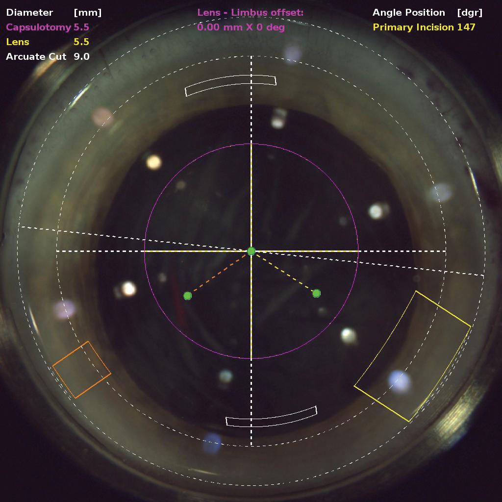
| The corneas of diabetic patients were found to take longer to recover after endothelial cell loss from FLACS, this study reported. Photo: Justin Schweitzer, OD. Click image to enlarge. |
Damage to the corneal endothelium during phacoemulsification occurs because of the ultrasonic energy used in the procedure. Effective phacoemulsification time and cumulative dissipated energy are both important risk factors for endothelial cell loss after cataract surgery. Studies suggest that femtosecond laser-assisted cataract surgery (FLACS) may result in less endothelial cell loss compared with phacoemulsification, as FLACS uses less energy to disrupt tissue.
A recent retrospective study compared endothelial cell loss after phaco and FLACS in patients with diabetes mellitus, a systemic disease that not only increases the risk of developing cataracts, but also affects the monolayer of corneal endothelial cells due to chronic metabolic changes at the cellular level. The researchers found that FLACS appeared to cause less damage than conventional phaco in these patients.
The study included 75 cataract patients (31 with diabetes) who underwent FLACS between August 2018 and November 2020. The same surgeon performed all cases.
The researchers reported no observed differences between groups regarding preoperative and intraoperative parameters, mean postoperative endothelial cell density, hexagonality and cell size. They noted that at one month, central corneal thickness was significantly greater in the diabetic group, but at three months, there was no significant difference between groups.
Overall, the researchers reported that changes in corneal endothelial cells between the two groups were comparable after FLACS. “The recovery of the cornea in patients with diabetes is longer than in normal controls,” the researchers wrote in their paper. “Despite good glycemic management, the corneal endothelium in diabetic patients is brittle to surgical trauma and has a weak ability to repair. The eyes of patients with diabetes are subject to various metabolic changes due to hyperglycemia; the aldose reductase in diabetic patients leads to the accumulation of polyols in cells, which act as an osmotic agent causing the swelling of endothelial cells.”
“Diabetes mellitus also reduces the activity of the Na+/K+ APTase in the corneal endothelium, which produces structural and functional changes in the cornea,” they added. “The diabetic endothelium was found to be under greater metabolic stress and had a less functional reserve after conventional phacoemulsification than the normal corneal endothelium.”
The researchers believe the lack of difference in corneal endothelial cell damage between the diabetic and nondiabetic patient groups may be due to the fact that FLACS uses less phacoemulsification energy, resulting in less damage to the cornea. “FLACS requires less phacoemulsification energy because the laser splits the nucleus,” they explained. “Because the endothelial cell loss correlates with the amount of energy used, FLACS reduces endothelial cell loss more than conventional phacoemulsification.”
Kang K, Song M, Kim K, et al. Corneal endothelial cell changes after femtosecond laser–assisted cataract surgery in diabetic and nondiabetic patients. Eye Contact Lens. 2021;1-6. |


