Ectasias can be difficult to diagnose, especially keratoconus (KC), which in some patients can progress slowly over several years. Recently, Chinese investigators wanted to clarify the relationship between corneal volume (CV) as measured with the Corvis ST device at different zones and corneal biomechanics in KC, along with the significance of CV in diagnosing and staging KC. Their prospective study included 456 KC eyes and 198 normal eyes as controls. The KC eyes were subdivided into groups by severity—mild, moderate or severe—based on corneal topography. CVs of the 3mm, 5mm and 7mm zones were obtained as were biomechanical parameters.
 |
|
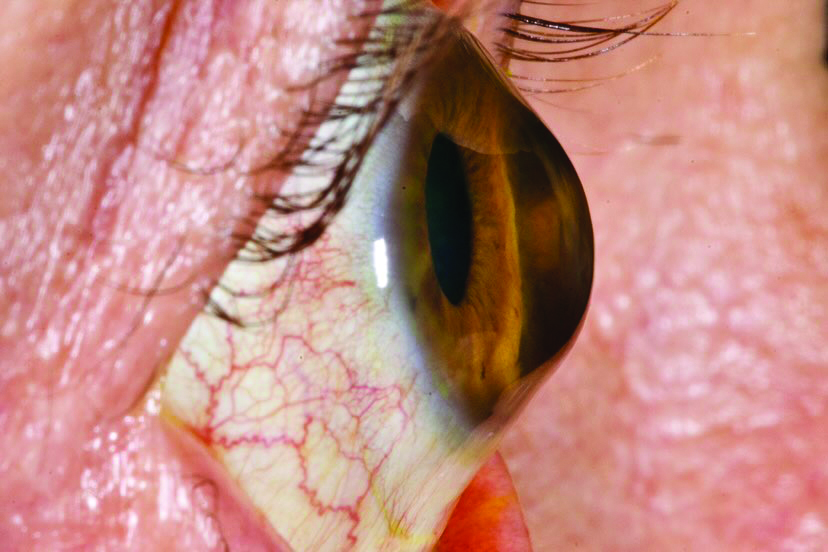
Progression of KC leads to corneal stroma destruction, which in turn causes instability of corneal biomechanical properties and weakened mechanical strength. Photo: Irving Martínez Navé, OD. Click image to enlarge. |
Results indicated that corneal volume of the 7mm zone displayed strongest correlation with many parameters: the first velocity of applanation, second applanation time, peak distance (width or bending distance), the maximal value of the ratio between the deformation amplitude at the apex and at 2mm from the corneal apex, the maximal value of the ratio between the deformation amplitude at the apex and at 1mm from the corneal apex, Ambrosio relational thickness to the horizontal profile, integrated radius, stiffness parameter at the first applanation and Corvis biomechanical index.
KC subgroups had lower CVs than controls for each diameter range, and there were differences seen between mild, moderate and severe subgroups for the 3mm zone. In this zone, CV displayed better diagnostic ability in each group to distinguish KC from normal corneas. The study authors relay that CV was shown to decrease in keratoconic eyes overall.
In their discussion, the authors elaborate that CV reflects topographical and pachymetric changes as well as characterizing corneal morphometric changes by doing so with one value. After controlling for central corneal thickness, correlations were seen between most biometric parameters and the CVs of both 7mm and 5mm zones. As the researchers explain, this confirms the relevance of the biometric and volumetric profile of the cornea in KC measurement of biomechanics.
Subsequently, “this implies that, among patients with KC, decreased CV values may indicate compromised corneal deformation. The progressive increase in corneal irregularities and a decrease in corneal CV might underlie the correlations between CV and biomechanical parameters,” the authors propose.
Clinically speaking, the researchers believe their results indicate “the CV of the 3mm zone has an acceptable diagnostic accuracy for KC detection and progression,” and that “the combination of CV and biomechanical analyses could enhance the ability to detect corneal ectasia and even to predict patients’ outcomes.”
Wu Z, Zhang Y, Li Y, et al. Correlation between corneal volume and corneal biomechanics and corneal volume significance in staging and diagnosing keratoconus. J Ophthalmol. May 29, 2024. [Epub ahead of print]. |