 |
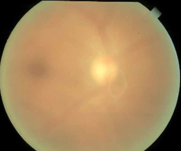
Look out for Weiss Ring when observing scans for progression. Photo: Joseph W. Sowka, OD. Click image to enlarge. |
Analysis of the retinal layer thickness is crucial for the diagnosis, treatment and follow-up of various ophthalmic diseases. Peripapillary retinal nerve fiber layer (pRNFL) thickness is one of the key considerations in glaucoma diagnosis and management, which also correlates with glaucoma severity. Therefore, the measurement of pRNFL thickness requires high reliability.
Researchers in South Korea believe that the Weiss ring, an annular vitreous opacity formed from peripapillary glial tissue that is frequently found in front of the optic disc, is one of the most frequent factors contributing vitreous opacity over the optic disc, which could affect pRNFL thickness measurements. Their study found a significantly thinner pRNFL in the inferior sector in eyes with a Weiss ring. The Weiss ring was commonly found in the inferior sector due to gravity, and its mobility led to the low reproducibility of pRNFL thickness measurements, especially in the inferior sector.
The team observed 205 eyes: 131 in the control group and 74 in which a Weiss ring was visible on OCT fundus images within a 3.46mm-diameter RNFL scan circle. The mean pRNFL thicknesses of the control and Weiss ring groups were 97.2µm and 94.6μm, respectively. In sectoral thickness, the inferior sector of the Weiss ring group was 112.1μm—significantly thinner than that of the control group (125.5μm).
The Weiss ring was located in 10 eyes (13.5%) in the superior sector, seven eyes (9.5%) in the temporal sector, 40 eyes (54.1%) in the inferior sector and 17 eyes (23.0%) in the nasal sector.
In analyses of reproducibility, the coefficient of variation and intraclass coefficient of the inferior sector measurement was 10.90% and 0.409, respectively, indicating low reliability of the measurement. The researchers said they believe the ring’s mobility could lead to the low reproducibility of pRNFL thickness measurements.
“After complete posterior vitreous detachment from the optic disc, a Weiss ring floating in the vitreous cavity above the optic disc is likely to sag downward due to gravity,” they wrote in their paper. “The inferior sector is a very important area when observing the trend in pRNFL thickness, especially in glaucoma patients, which would be affected by measurement errors from the Weiss ring.”
The study concluded that physicians should consider these impacts of a Weiss ring when interpreting changes in pRNFL thickness. When the Weiss ring was found to be on the 3.46mm-diameter scan circle in the RNFL deviation map, the measurement was likely to be unreliable and repeated measurements should be considered.
Lee MW, Kim MS, Yu HY, et al. The Weiss ring, a major confounding factor for measurements of peripapillary retinal nerve fiber layer thickness: the impact of the Weiss ring on RNFL thickness measurements. Am J Ophthalmol. January 13, 2022. [Epub ahead of print]. |