 |
|
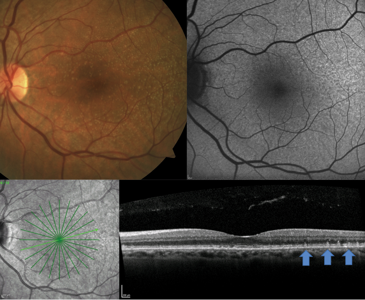
The updated severity scale fits modern definitions of late AMD, which include geographic atrophy, and provides increased prognostic accuracy by incorporating reticular pseuodrusen (shown). Photo: Jessica Haynes, OD. Click image to enlarge. |
Estimating a patient’s risk for progression to late AMD in the clinic helps with counseling, making treatment plans and justifying home monitoring and interventions. Of the several risk calculators available, such as the AREDS 9-Step Severity Scale and the Advanced AMD Risk Calculator, the AREDS Simplified Severity Scale was designed for efficient clinical use, with easy to memorize risk categories based on simple grading. However, experts pointed out in a recent Ophthalmology report that these approaches focus on only two macular features: soft drusen and pigmentary abnormalities. Now that the roles of non-central geographic atrophy (GA) and reticular pseudodrusen (RPD) in AMD progression are recognized, researchers proposed an update to the AREDS Simplified Severity Scale. Their new scale demonstrates greater predictive accuracy while remaining easy to use.
The post-hoc analysis of the AREDS (n=2,719) and AREDS2 (n=1,472) cohorts included participants with no late AMD at baseline. In the AREDS, the five-year progression rates to late AMD, based on the Simplified Severity Scale (levels 0 to 4) were 0.3%, 4.5%, 12.9%, 32.2%, and 55.6%, respectively. In the present study, the researchers calculated the five-year progression rates with two updates:
- Non-central GA considered part of the outcome instead of only a risk feature.
- Scale separation according to RPD status (via color fundus photographs graded by a validated deep learning algorithm).
With these two updates, the proportion of eyes progressing to late AMD by year five was 8.4% in those without RPD and 40.6% in those with RPD. The revised Simplified Severity Scale showed that five-year progression rates for levels 0 to 4 were as follows:
Rate of Progression to Late AMD at Five Years
# of Risk Factors | RPD Absent | RPD Present |
0 | 0.3% | 2.8% |
1 | 4.3% | 8.0% |
2 | 11.6% | 29.0% |
3 | 26.7% | 58.7% |
4 | 50.0% | 72.2% |
External validation using the AREDS2 (levels 2 to 4) demonstrated similar progression rates: 15.0%, 27.7% and 45.7% without RPD, and 26.2%, 46%, and 73% with RPD, supporting this scale’s generalizability.
Since the new scale for patients without RPD is very similar to the original, “physicians can continue to use these values in the setting of RPD absence,” the researchers wrote in their Ophthalmology paper. For RPD presence, they wrote, “physicians could either use these values themselves or consider that the risk is approximately double for the three middle levels, but higher for level 0 and lower for level 4.”
Agrón E, Domalpally A, Chen Q, et al. An updated simplified severity scale for age-related macular degeneration, incorporating reticular pseudodrusen: Age-Related Eye Disease Study Report No. 42. Ophthalmology 2024. [Epub ahead of print]. |