The assessment of retinal volume is an important parameter in understanding ocular health and detecting countless potential abnormalities. Changes in retinal volume—which some clinicians are now able to capture using widefield swept-source optical coherence tomography (SS-OCT)—can provide valuable insights into the progression of ocular diseases and guide treatment decisions. Therefore, optometrists need to have a comprehensive understanding of the factors influencing retinal volume, in healthy cases for comparison in addition to various disease states. To provide some of this knowledge, a recent study assessed retinal volume in individuals without ocular disorders using widefield SS-OCT and investigated its association with demographic and anatomical characteristics.
 |
|
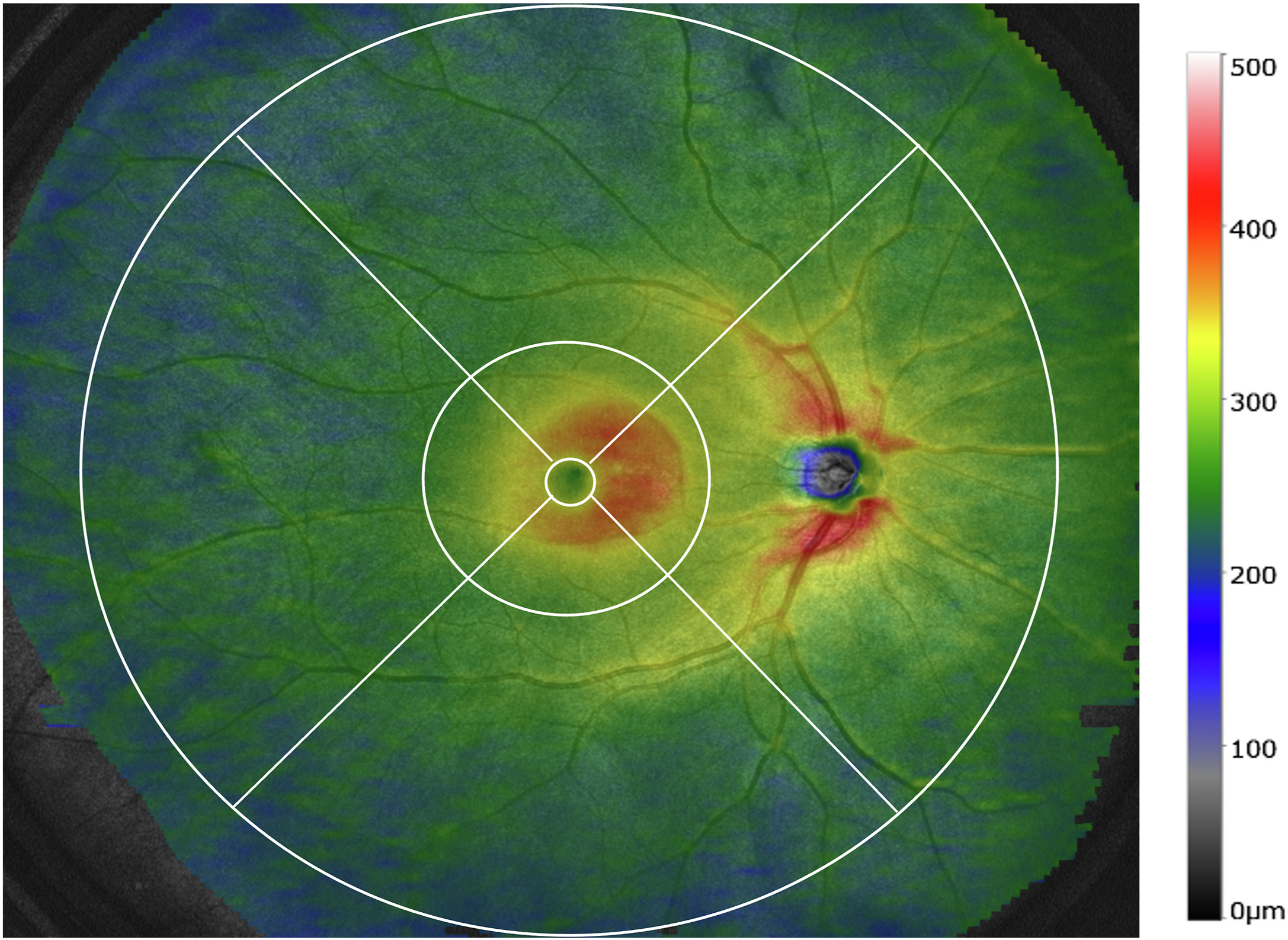
Researchers observed a significant association between age and axial length and macular retinal volume, as well as between laterality and peripheral retinal volume. This color-coded en face retinal thickness map from the study averages the scans of healthy volunteers. The circles are at diameters of 1mm, 6mm and 20mm from the fovea. Photo: Chiku Y, et al. Ophthal Sci. June 21, 2024. Click image to enlarge. |
The observational, cross-sectional study involved 332 eyes of 166 healthy participants, including 101 women and 65 men, with a mean age of 43 years. Eyes were imaged with OCT-S1 (Canon) using a protocol centered on the fovea cube scans (20x23mm) of SS-OCT images. Retinal volume was evaluated in different regions, including the macula and peripheral retina, as shown in the photo, which is a composite of the study data for all subjects.
In the macular region—shown here in the 6mm diameter middle ring of the OCT image—the nasal quadrant had the greater retinal volume (2.233mm3), followed by the superior (2.137mm3), inferior (2.057mm3) and temporal (2.034mm3) ones. In the peripheral retina—the larger, outer ring extending from 6mm outward to 20mm from the fovea—the nasal quadrant had the highest volume (18.4mm3), followed by the superior (16.4mm3), temporal (15.5mm3) and inferior (15.2mm3) quadrants.
The multivariate analysis showed that older age and longer axial length were associated with smaller macular retinal volume, while older age and left eye were associated with smaller peripheral retinal volume. In their paper on the study, published in Ophthalmology Science, the authors wrote that “these results suggest that age and axial length are important factors in the assessment of retinal volume.”
The link between retinal volume and age has been indicated in previous studies reporting on retinal layer thickness across age groups. One that the authors of the present study pointed to in their paper concluded that “the thicknesses of the RNFL, ganglion cell layer, inner plexiform layer, inner nuclear layer and photoreceptor inner segment were negatively correlated with age.”
Although this study found the peripheral retina to be significantly smaller in the left eye than in the right eye, the authors advise that these results be interpreted with caution. “In our examination protocol, we first examined the right eye, which may have affected our results,” they explained in their paper. “That is, the patient may have lost concentration, and the image quality may have been affected by eye or head movements when the left eye was examined.” Additionally, “previous studies on choroidal thickness and retinal ganglion cell layer thickness have not reported any differences between the left and right eyes.”
Regional differences in retinal thickness were also observed within the macula and peripheral retina; the temporal area of the macula was significantly smaller than all other areas (1mm to 6mm) whereas the inferior area was significantly smaller than all other areas in the peripheral retina (6mm to 20mm). The authors pointed out that the former finding is consistent with that of a previous study showing that the thinnest retinas are temporally located in the macula.
In summary, this research provided two main conclusions about retinal volume in healthy eyes as assessed on widefield SS-OCT: (1) age and left eye are negatively correlated with peripheral retinal volume, and (2) the thinnest part of the retinal quadrant differs between the macular and peripheral retinas.
Chiku Y, Hirano T, Hoshiyama K, et al. Assessment of retinal volume in individuals without ocular disorders based on widefield swept-source optical coherence tomography. Ophthalmol Sci. June 21, 2024. [Epub ahead of print]. |