 |
| Corneal thickness is affected by an individual's age, gender and height, study shows. Photo: Maria Walker, OD. |
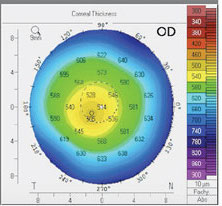
Central and peripheral corneal thickness are important parameters that give insight into the structure’s health and function, but there is little data characterizing the increase in corneal thickness from central to peripheral. The aim of a recent study was to report on these cornea characteristics and investigate associated factors in a population-based setting.
The Gutenberg Health Study is a prospective, population-based research effort that examined 9,729 participants aged 40 to 80 during a five-year follow-up using Scheimpflug imaging. Corneal thickness was assessed in each participant at the apex, as well as in the corneal center and in rings with 2mm, 4mm, 6mm, 8mm and 10mm diameters around the corneal center. The increase in corneal thickness toward the periphery was also evaluated.
The team found that older age is associated with a thinner central and peripheral cornea. Age-related changes of the cornea were also reported for different corneal layers by various studies.
“The present study provides evidence that higher intraocular pressure is associated with a thicker cornea in the center and periphery,” the authors noted in their paper, published in Acta Ophthalmologica.
When analyzing the distribution of corneal thickness with respect to quadrants, the superior cornea was the thickest, followed by the nasal and the inferior regions; the temporal quadrant was the thinnest.
The authors also observed that a thicker central and mid-peripheral cornea (until 6mm) was associated with male gender, which is similar to findings from previous studies, and that body height was associated with a thinner cornea in the corneal periphery.
The corneal thickness at the 10mm ring showed an inverse association with hyperopic refractive error, which was not present in the central cornea.
“White-to-white distance, corneal radius and refractive error are linked to corneal thickness in the peripheral cornea,” the authors explained. “Furthermore, we observed an increased thickness of the 8mm and 10mm circles with pseudophakia. One reason for this association might be the scar after clear corneal incision.”
In contrast to these results, “Glaucoma was linked to a thinner cornea at the 4mm circle and toward the periphery independently of intraocular pressure,” the authors noted. “This new information is particularly of clinical importance as peripheral corneal thickness is important for surgical procedures like laser or refractive surgery, glaucoma assessment and observation of corneal diseases such as keratoconus and Fuchs’ endothelial dystrophy.”
Rahmani M, Ortiz-Toquero S, Martin R. Referral pattern and co-management of keratoconus patients in primary eye care: a survey of three European countries. Cont Lens Anterior Eye. November 10, 2021. [Epub ahead of print]. |