iCare One Personal Tonometer
Designed for glaucoma patients who need regular IOP monitoring, the iCare One uses a rebound method of measurement for at-home testing. Avery light probe makes momentary, gentle contact with the cornea and does not require anesthetic drops. The iCare tonometer is already used in U.S. doctors’ offices, but this at-home version is currently awaiting FDA approval.
 A recent study at Duke University found the test was easy to learn, well tolerated, accurate and reliable.1 It includes two adjustable support elements and an eye cup for easy self-tonometry, but also can be administered by health professionals and even inexperienced users, the manufacturer says. Its indicator displays 11 different pressure zones—ranging from 5mm Hg to 50mm Hg. Featuring a built-in inclination sensor that can correct results automatically, the iCare One can be set to take just one measurement at a time or a series of six measurements.
“The iCare One is interesting because the unit can be attached to a PC via USB and generates IOP curves from stored data,” says Kristopher May, O.D., of Coldwater Vision Center in Mississippi. “It lets you know how the patient’s really doing and can alert you if there are any abnormal results.”
Diaton Tonometer
The Diaton Tonometer provides reliable measurements and makes it possible to diagnose glaucoma in its early stages, allowing you to devise an appropriate care and treatment plan. “When I first saw a Diaton Tonometer, I assumed it was just another tono-pen, but when I saw it used, I was impressed,” Dr. May says. “It’s actually a transpalpebral tonometer—it takes IOP through the lid and measures against the superior sclera.” 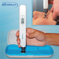
It’s effective in obtaining IOP measurements for many challenging patient populations, including: those with chronic conjunctivitis, erosions, edema and corneal dimness; people who have undergone corneal surgeries; and immobilized patients and children. Also, patients don’t need to remove their contact lenses.
“Since the device does not touch the cornea, there is little risk of corneal abrasion or decreasing visual acuity,” Dr. May says. “Additionally, there is no dependence on corneal pachymetry.” Sterilization and anesthesia are not required, and the measurement range spans from 5mm Hg to 60mm Hg. A single measurement takes no longer than three seconds, and one set of batteries provides at least 1,500 measurements, according to the manufacturer.
iWellnessExam with iVue SD-OCT
A recent addition to Optovue’s iVue portable spectral domain OCT, the iWellnessExam offers a quick, noninvasive test to complement your routine comprehensive eye exam. “The iWellness Exam is a very simple procedure to perform and to interpret,” says Jerome Sherman, O.D., a faculty member at the SUNY College of Optometry in New York. “It has the ability to detect problems of the central retina and problems of the optic nerve simultaneously.”
iWellness images provide a cross-sectional view of the retinal layers with 5µ resolution, as well as a retinal thickness map and a ganglion cell complex map. In fact, Dr. Sherman recently used it to screen a patient with a normal-looking fundus, and it revealed a retinal problem and a ganglion cell abnormality within seconds. 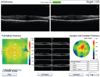
Early detection enables early intervention, which could prevent or slow vision loss. The iWellness exam is recommended annually regardless of symptoms, allowing you to observe subtle changes over time or to confirm the patient’s ocular health. Dr. Sherman suggests that, one day, perhaps it could become as routine as annual dental X-rays. “It gathers so much information in such a short period of time,” he says. “And it’s completely noninvasive—there’s nothing dangerous whatsoever about it. It is certainly possible that SD-OCT will soon become the standard of care.”
EyeQuick Digital Ophthalmoscope Camera
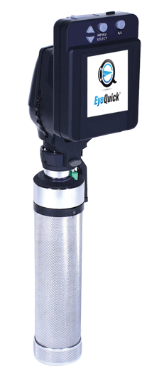
The new FDA-approved EyeQuick was designed to combine the portability of an opthalmoscope, the imaging power of a tabletop retina camera and the ease of use of a commercial digital camera. This wireless ocular imaging system captures pictures and videos of the anterior segment and retina. It also allows you to review images instantly on the built-in LCD screen.
“Even physicians who have a retinal camera will be able to enhance patient care through the EyeQuick’s portability and versatility,” says inventor Marc Ellman, M.D., of the Southwest Eye Institute in El Paso, Texas. While traditional retinal cameras can cost $20,000 to $40,000, the EyeQuick offers a quick return on investment with a price tag of about $6,000. When appropriate, doctors can use it to bill for retinal and anterior segment photographs.
“Now this is an eye-dork toy! It’s portable, small, simple and takes surprisingly good images and videos,” Dr. May says. “You can upload via USB to your EMR or to your computer, which makes it perfect for nursing home or satellite clinics.”
UBM Plus Portable Imaging Device
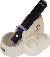 With a 48MHz probe that plugs directly into a laptop or desktop computer, Accutome’s UBM Plus portable anterior segment imaging device can be used in almost any setting. It features data analysis tools for measuring sulcus-to-sulcus, anterior chamber depth, positioning of intraocular lenses and the filtration angle of the eye. Its all-in-one probe is designed to eliminate signal loss and provide sharp images.
“Ultrasound biomicroscopes are great for anterior chamber angles and iris lesions,” Dr. May says. "Because the UBM Plus software is upgradeable, it does help to offset the initial cost in the long run. “If I’m buying a big-ticket item, I’m looking at upgradeability and its longevity,” Dr. May says. “I’ve got to be able to expand it, to do other testing with it and to update it so it can stay current.”
The advanced software captures 34-second film loops. While reviewing the scans, you can simply take a snapshot of a particular image. The built-in reports template pulls these images together with collected information to create a detailed report in seconds. Because the UBM Plus interfaces easily with electronic medical records, it makes sharing this information with other doctors simple.
OPD-Scan III 3D Wavefront System
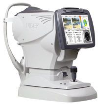 The OPD-Scan III 3D Wavefront system from Nidek and Marco is a refractive power/corneal analyzer that can evaluate a patient’s total visual system. It combines pupillometry, autorefraction/keratometry, corneal topography, optical path difference (OPD) and wavefront analysis into one diagnostic workstation.
New features include ARK function automation, wavefront visual acuity maps, a full 9.0mm measurement area, CT blue light and electronic medical record integration. In 10 seconds, OPD-Scan III can capture: the spherical aberration of the cornea for aspheric intraocular lens selection; lenticular—residual astigmatism; angle kappa; pre/post toric intraocular lens measurements; corneal pathologies; mesopic/photopic pupil size; retro illumination image; Zernike graphs; corneal refractive power map; and intraocular lens tilt or decentration.
“The newest OPD provides just about everything you ever wanted to know about the cornea in one scan from one device,” Dr. May says. The only things that might improve it, he says, would be the addition of more novel analytics like tear film data and output to a prescription lens system.
Nova-DN VEP Vision Testing System
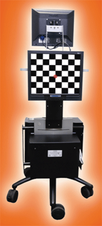 The Diopsys NOVA-DN uses short duration transient Visual Evoked Potential technology (SD-tVEP) to record the electrical responses of a patient’s entire vision system. It provides easy-to-read reports that give the doctor a simple way to evaluate optic nerve function and optic nerve disease, including glaucoma and other neurovisual disorders.
“It’s a way of looking at the entire pathway of vision,” Dr. Sherman says. “It’s a completely objective test; hence, it can be used for infants, children, and patients with very subtle abnormalities that we may not be able to pick up with standard tests.”
The test takes between four and six minutes, and doctors are able to compare tests over time to track disease progression. Clinicians may use VEP in addition to traditional testing methods to enhance diagnosis and treatment of functional disorders.
A recent paper in the Journal of Glaucoma showed it to be a fast, objective method to screen for functional damage in glaucomatous eyes, and Dr. Sherman believes it will also prove to be extremely useful in assessing patients who have loss of function from conditions such as multiple sclerosis or other central nervous system disorders.2
Foresee PHP: the Home Version
Foresee, an FDA-cleared, teleconnected, home-based system from Notal Vision, enables frequent monitoring of age-related macular degeneration between eye exams. This home monitoring program uses preferential hyperacuity perimetry (PHP) testing to detect central and paracental metamorphopsia, allowing you to detect important visual changes even before noticeable symptoms appear.
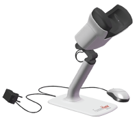 “The home device works just like the office device except that it’s something the patient can take home and test themselves,” Dr. Sherman explains. “Early detection in wet macular degeneration is crucial, and this device allows us to do that.” The system sends results to a reading center, where they are interpreted to see if there have been any changes or abnormalities.
The Foresee Home PHP provides helpful monitoring data, but it is not intended to diagnose. If the patient needs additional testing in-office, the Notal Vision test center will alert you. “We’ll still see this patient in the office three to four times a year, but now we can see them every day in a sense because the lab will be monitoring them on a daily basis,” Dr. Sherman says.
Macular Integrity Assessment Tool
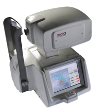 The MAIA from Ellex is a scanning laser ophthalmoscope with an eye tracker and microperimeter. It allows you to assess, evaluate and monitor changes in average macular threshold sensitivity in AMD patients. “The major advantage is that it allows us to measure the sensitivity in decibels of specific points across the central retina with extreme accuracy,” Dr. Sherman says. “It’s possible to show that you can measure the sensitivity of individual drusen within the retina.”
The microperimeter can run any of three tests on the central 10 degrees of the retina:⎯fast supra‐threshold analysis, detailed threshold or follow-up threshold analysis. MAIA also includes threshold sensitivity analysis software, which allows you to process the measured data and evaluate macular function as compared with a reference database of 270 normal subjects.
“It is different than any other visual field system available at the present time because of the automated eye tracking,” Dr. Sherman notes. “If the patient is not looking in the appropriate direction, it automatically can put the stimulus where it belongs on the retina.”
Zeiss i.ProfilerPlus System
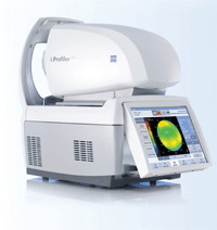 The i.ProfilerPlus combines an autorefractor, wavefront aberrometer and an Atlas 9000 corneal topographer into one unit that can measure both eyes automatically in less than one minute. “The i.ProfilerPlus links to Zeiss i.Scription technology, which utilizes Zeiss VoluMetric merit function to combine the autorefraction, aberration data and the doctor’s subjective refraction,” Dr. May explains. “The result is a super accurate prescription that is then custom-manufactured into a free form spectacle lens.”
The i.ProfilerPlus measures the distribution of the refractive power across the whole pupil, permitting more accurate calculation of the entire refractive status of the eye. These measurements can also be presented graphically in an extensive array of contour maps and vision simulations.
It’s useful in a variety of clinical applications: evaluating the complete refractive status of the eye; fitting soft and rigid contact lenses; monitoring ocular disease processes; and managing or comanaging refractive and surgical interventions. The process is fully automatic and can easily be performed by staff with minimal training.
With this type of advanced technology, optometrists can refocus their attention on improving vision. “Eye care got here because we helped people see better, which gave us the opportunity to care for their ocular health,” Dr. May says. “To use a slightly more country expression, ‘You dance with the one that brung you.’”
1. Flemmons MS, Hsiao YC, Dzau J, et al. Home tonometry for management of pediatric glaucoma. Am J Ophthalmol. 2011 Jun 18. [Epub ahead of print].
2. Prata RS, Lima VC, De Moraes CG, et al. Short duration transient visual evoked potentials in glaucomatous eyes. J Glaucoma. 2011 May 10. [Epub ahead of print].

