 |
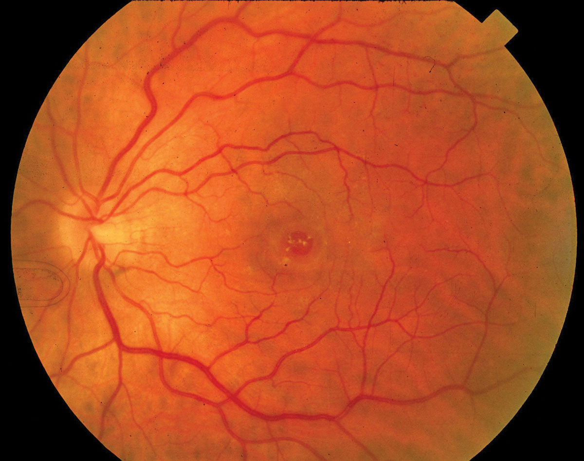
| At the onset of a full-thickness macular hole, a foveal inner tear is more likely to occur on the temporal side of the macular hole or the side with the highest posterior vitreous separation height. Photo: Diana Shechtman, OD. Click image to enlarge. |
With the advent of OCT, analysis of the retina in greater detail has been possible, leading researchers to better understand the mechanisms behind macular hole development, vitreous detachment and other changes to the foveal structure. Despite this, the mechanism of macular hole progression is poorly understood.
Stage-one macular holes often resolve spontaneously, while stages two to four may require surgical repair. Differentiating between stages one and two is key when deciding whether to perform surgery. To better characterize macular hole progression, researchers examined the relationship with posterior vitreous detachment. They hypothesized that macular hole formation may occur as a “byproduct” of the posterior vitreous detachment processes and identified a possible predictive factor.
The retrospective study included 742 patients with full-thickness macular hole or impending macular hole stages one to three, defined as posterior vitreous membrane and vitreoretinal adhesion size ≤1,500µm. If a patient’s contralateral eye demonstrated the focal type of vitreomacular adhesion (≤1,500µm), it was also included in the analysis.
On OCT, the researchers analyzed patients’ posterior vitreous separation height (PVSH), the distance between the posterior vitreous membrane and the surface of the retina, at 1mm from the center of the macular hole or fovea. Then, they calculated several trends among the PVSH in each of the four directions (nasal, temporal, superior and inferior). They reported the following in each of the four directions: vitreomacular adhesion < macular hole stage one = macular hole stage two < macular hole stage three.
In their paper, the researchers described the initial stage-two macular hole (onset of full-thickness macular hole) as “the presence of a gap in only one of the four directions from the center of the macular hole.”
They observed that the greater the PVSH, the greater the chances of this gap occurring in stage-two eyes. Additionally, a temporal gap was significantly more likely to occur than a nasal gap.
“Patients with a large PVSH are at high risk of developing macular hole stage two,” they wrote, but “unexpectedly, progression from macular hole stage one to stage two was observed in the absence of significant changes in PVSH.”
The odds of developing this stage-two full-thickness hole were also reportedly higher among females than males for a PVSH greater than 300µm. “Ophthalmologists must be aware that as PVSH height increases, the odds ratio of progression to macular hole stage two increases more in female than in male patients.”
Sakaguchi H, Kabata D, Sakimoto S, et al. Relationship between full-thickness macular hole onset and posterior vitreous detachment: a temporal onset theory. Ophthalmol Sci. May 2023. [Epub ahead of print]. |
