A limbal relaxing incision is commonly combined with a toric IOL when the need for astigmatism correction exceeds the toricity of currently available IOLs.
Astigmatic laser refractive surgery has become accessible only years after myopic treatments had been around. With current cataract surgery, postoperative astigmatism has become a cringe-worthy event. In the era of small-incision cataract surgery—where good uncorrected acuities are not only routine, but expected by patients—residual refractive error is considered (by patients, at least) a negative outcome.
Unfortunately, postoperative astigmatism isn’t an unusual event. In one very large study of a cataract population, corneal astigmatism between 0.75D and 1.50D was present in more than 40% of patients, with higher levels in nearly 20%.1 Historically, this figure has dovetailed poorly with our ability to treat astigmatism. Intraocular lens technology has been primarily spherical in its refractive properties, and until recently correction of corneal astigmatism has been an after-thought based on wound positioning and corneal incisional surgery. As technology has increased, however, so too has our ability to treat astigmatism.
So, in this age of baby boomers with both cataracts and astigmatism, what are their options—and what does the science say about them?
Limbal Relaxing Incisions
For years, our go-to option for expected post-cataract surgery astigmatism has been the limbal relaxing incision, or LRI.
LRIs are based on the same principle as all-incisional refractive surgery; that is, a partial-thickness cornea incision leads to flattening of the cornea—a premise that dates back to Dr. Herman Snellen in the mid-1800s.2 Technically, LRI is a tissue addition procedure (like other incisional refractive surgeries); as the gaped wound heals, it fills with scar tissue and results in a flattening of that meridian. The surgeon makes an arcuate or tangential partial thickness incision just onto the clear cornea. The incision is along the steep axis of astigmatism, with its depth and length determined by the amount of astigmatism to be treated; a longer (up to 90 degrees) and deeper cut leads to greater dioptric flattening.
By comparison, in astigmatic keratotomy (an older procedure), the incisions were placed within the central corneal zone, which led to greater refractive impact per incision size, but also greater refractive instability.
The reason for an LRI performed during a cataract operation is to correct any astigmatism that exists at the corneal plane, as IOLs have traditionally utilized spherical or aspheric optics. The procedure can be done along with the cataract while on the operating room table, and it offers a low risk, cost-effective means of minimizing expected postoperative astigmatism.
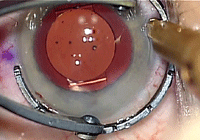
Newer-generation toric IOLs are designed for better stability and less rotation than previous toric implants. They’re also coming out in greater ranges of astigmatic correction. Photo: Walter Whitley, O.D.
The strengths of LRI are its simplicity and ease of use: It’s a comparatively non-invasive procedure that can safely correct corneal astigmatism, and the flexibility of incision placement allows the correction of some amount of irregular astigmatism as well. For example, we recently had a 41-year-old male in our clinic for a LASIK evaluation. He saw 20/30 O.U. uncorrected, and his only refractive error was 1.50D of simple astigmatism. Instead of LASIK, we suggested a less expensive, less invasive LRI first. The patient agreed and the surgery soon took place. At his one-month follow-up, the patient was 20/15 and thrilled to have saved some of the cost of surgery. In short, LRI does maintain its place in clinical and surgical practice.
More commonly, we combine an LRI with a toric IOL for an astigmatism correction that exceeds the toricity of currently available IOLs (a magnitude that seems to be ever expanding).
The weakness of LRI is the procedure’s unpredictable nature. Despite well-known nomograms for incisional depth and length for refractive effect, it is not a 100% accurate or consistent response. LRI is described quite succinctly as “more art than science,” says Robert Osher, M.D., medical director at Cincinnati Eye Institute.3
Unfortunately, it’s not rare to have minimal impact on refractive cylinder with LRI, despite following the nomograms. In one study of LRI paired with cataract surgery in 55 eyes, the mean preoperative corneal astigmatism was 1.90D, which was reduced to 1.00D by six months post-op.4
A study of the effects of LRI for mixed corneal astigmatism showed a similar result with roughly 3.30D of preoperative cylinder, which was reduced to 1.60D postoperatively.5 This lack of predictability and accuracy has created the niche for a more consistent means of correcting astigmatism, particularly in conjunction with cataract surgery.
This is where toric IOLs are gaining ground.
Toric IOLs
In theory, an implanted toric lens that has good capsular support should be stable within the eye and provide correction of astigmatism. Optically, the magnitude of this correction will be about 70% the toricity of the lens. Said differently, an IOL with 3.00D of toricity will correct about 2.00D of corneal astigmatism.
One retrospective study compared the theoretical benefit of a toric IOL vs. a spherical IOL and LRI.6 Of the patients who received the toric IOL, mean uncorrected visual acuity averaged 20/30, while those who received the mixed procedure achieved 20/40 vision on average. As many as 70% of toric IOL patients saw 20/30 or better, compared to 51% of those receiving LRIs.
| LRI Revisited: LenSx and LRI | ||
The LenSx is a femtosecond laser instrument that, rather than replacing the surgeon, assists with some key features of surgery, including creation of clear corneal incisions, creation of the capsulorhexis, and lens fragmentation. Stabilization of the globe, completion of phacoemulsification, removal of the lens particles, and IOL implantation are still in the hands of the surgeon. One of the interesting features of the LenSx––in relation to corneal astigmatism and cataract surgery––is its ability to use the laser to also create LRI incisions. The laser uses a novel nomogram to determine incision magnitude. “The first thing that struck me about the LRIs generated with LenSx is how clean the incisions look,” says David Coulson, O.D., of Barnet Dulaney Perkins Eye Centers, in Arizona, whose practice utilizes the LenSx. “Secondly, the incisions are a little more central, coming a couple millimeters in from the limbus. This makes them a bit like a hybrid LRI/astigmatic keratotomy.” Time will tell if laser-generated LRIs are more stable or predictable than the bladed variety. But, according to Dr. Coulson, results so far have been encouraging. “It does seem like the predictability of the incision and level of control with it has enhanced outcomes,” he says. Still, Dr. Coulson points out that this technology is supplemental for postoperative astigmatism and doesn’t replace toric lenses. “We use the technology to treat lower levels of corneal astigmatism. When the toricity gets to 1.25D [and above], we still recommend the toric IOL options.” |

However, one of the drawbacks of the toric lens used in this study, STAAR Toric IOL (STAAR Surgical), was rotational instability.6 About 18% of patients showed rotation between 20 to 40 degrees, and 7% had greater than 40 degrees of rotation. All IOLs were able to be repositioned, but the result was illustrative of the problems that occurred with older-generation toric IOLs.
While older toric lenses were an improvement over LRIs, they also unfortunately suffered from issues of stability and predictability—the premise upon which they were based.6,7
This rotational instability is significant because toric lenses lose 3.3% of their astigmatic correction for each degree of misorientation. At 30 degrees net misrotation, there is no net astigmatic correction. At 30 degrees off axis, the lens actually creates refractive astigmatism.
The newer generation AcrySof IQ Toric IOL (Alcon Surgical) was designed to minimize these shortcomings. The lens shares a similar design with the AcrySof Single-Piece IOL, a lens that was designed with stability-enhancing features such as more stable haptics and a bio-adhesive polymer base. While such features may sound like creative marketing, these modifications actually seem to work. For example, Ed Holland, M.D., of the Cincinnati Eye Institute and one of the world’s foremost anterior segment specialists, conducted a study to assess stability and visual outcomes with this IOL.7 The study results showed good stability with minimal rotation (a mean of 3.8 degrees).
Of the 256 implants in this study, only three rotated by more than 15 degrees over the year of follow-up. Two-thirds (65%) of the patients who received the toric IOL had uncorrected acuity of 20/25 or better, compared to 29% of the controls who received the AcrySof spherical lens.8
Another study on bilateral AcrySof Toric implants showed axis alignment within five degrees in 91% of patients and within 10 degrees in 99% of patients.9 This equated to good visual outcomes for patients—uncorrected acuities of 20/40 or better in 99% of patients, and 20/20 or better in 60%.
Also, it’s helpful to have implants that come in a range of corrections. There are seven different AcrySof Toric lens options correcting corneal cylinder from 1.00D through more than 4.00D.
Of the different treatment options available, “I prefer the toric IOL options to LRI. I’ve found their predictability to be superior to that of LRIs,” says Douglas Devries, O.D., co-owner of Eye Care Associates of Nevada, a large surgery practice. “That’s not to say I never recommend LRIs. Sensitive patients with less than 0.75D of cylinder—those who fall below the scope of the toric IOL—are fine candidates.”
Dr. Devries also considers LRI for astigmats with cylinder beyond the scope of correction with the lens. “In these cases, we’ll use a combined toric IOL/LRI procedure,” he says.
Comanagement Care
From a referring optometrist’s standpoint, there is very little additional data that needs to be gathered at the preoperative exam to utilize the toric lens. In order to determine if astigmatism will be present after surgery, manual or topographic keratometries could be performed; however, this will be repeated at the surgery center.
Discuss toric IOLs with all patients who have refractive astigmatism. In most cases, this is a fair indication if there will be residual cylinder after surgery. If a referring optometrist doesn’t bring up the different IOL options with their patient prior to cataract surgery, the O.D. is essentially relinquishing any advisory role in the process to the surgeon.
Toric IOL or LRI: What Do Surgeons Prefer?
When faced with a patient with astigmatism, 57% of surgeons say they prefer to use toric IOLs, according to a survey in Review of Ophthalmology.
Thirty-one percent like limbal relaxing incisions, 7% place their entry incision on-axis, and 5% use a post-op refractive procedure to handle the astigmatism.
This discussion doesn’t need to be exhaustive—it’s sufficient to simply make the patient aware of the lens options available and offer any professional preference you have.
Postoperative follow-up for toric lenses is only slightly more technical than with standard IOLs or LRIs. In addition to the normal postoperative exam, toric lenses require evaluation of lens orientation under dilation. If the lens is rotated significantly (such that the patient appreciates the difference), a surgical repositioning is in order.
Early rotation (rotation that occurs in the first postoperative month) is much more common than late rotation.6-8 This often coincides with typical postoperative follow-up because most three- or four-week follow-ups involve dilation and allow assessment of lens position.
Repositioning of the lens is an effective means of treating the problem—but the timing of it is important.6 “Referring O.D.s need to know the intended axis of rotation and evaluate it closely,” Dr. Devries says. “It’s very important to understand that the critical time for repositioning of these lenses is within the first month. Repositioning [during this period] is more effective and leads to greater stability in the long run.”
There is a definite evolution going on within our cataract patient base. The baby boomer population views cataract surgery not only as a means to see better with correction, but also as a refractive procedure in itself. To meet this demand, reliable IOL technology has been developed to aid in the neutralization of postoperative astigmatism. With no additional drawbacks outside of occasional rotation issues and cost passed onto the patient, toric IOLs are also very safe devices to use.
In summary, “One of the keys to postoperative success—and we hear this all the time with multifocal IOLs—is managing and meeting patient expectations. With astigmatic corrections, it’s no different,” Dr. Devries says. “The toric lenses give the patient a better chance of achieving the outcome I discussed with them. This makes me look good and it makes for a happy patient.”
Dr. Bronner is in practice at Hollingshead Eye Center, a secondary care center in Boise, Idaho. He has no financial interest in any of the products or companies mentioned.
1. Hoffer KJ. Biometry of 7,500 cataractous eyes. Am J Ophthalmol. 1980 Sep;90(3):360-8.
2. Chalita MR, Krueger RR, Boxer-Wachler B. Refractive Aspects of Cataract Surgery. In: Yanoff M, Duker JS, eds. Ophthalmology. 2nd ed. Philadelphia: Mosby Inc; 2004:228-37.
3. Osher R, et al. Astigmatism Correcting IOLs: Pearls for Your Practice. Ocular Surgery News (suppl). 2009:1-15.
4. Carvalho MJ, Suzuki SH, Freitas LL, et al. Limbal relaxing incisions to correct corneal astigmatism during phacoemulsification. J Refract Surg. 2007 May;23(5):499-504.
5. Bayramlar HH, Dağlioğlu MC, Borazan M. Limbal relaxing incisions for primary mixed astigmatism and mixed astigmatism after cataract surgery. J Cataract Refract Surg. 2003 Apr;29(4):723-8.
6. Sun XY, Vicary D, Montgomery P, Griffiths M. Toric intraocular lenses for correcting astigmatism in 130 eyes. Ophthalmology. 2000 Sep;107(9):1776-81; discussion 1781-2.
7. Till JS, Yoder PR Jr, Wilcox TK, Spielman JL. Toric intraocular lens implantation: 100 consecutive cases. J Cataract Refract Surg. 2002 Feb;28(2):295-301.
8. Holland E, Lane S, Horn JD, et al. The AcrySof Toric intraocular lens in subjects with cataracts and corneal astigmatism: a randomized, subject-masked, parallel-group, 1-year study. Ophthalmology. 2010 Nov;117(11):2104-11.
9. Ahmed II, Rocha G, Slomovic AR, et al; Canadian Toric Study Group. Visual function and patient experience after bilateral implantation of toric intraocular lenses. J Cataract Refract Surg. 2010 Apr;36(4):609-16.
