 |
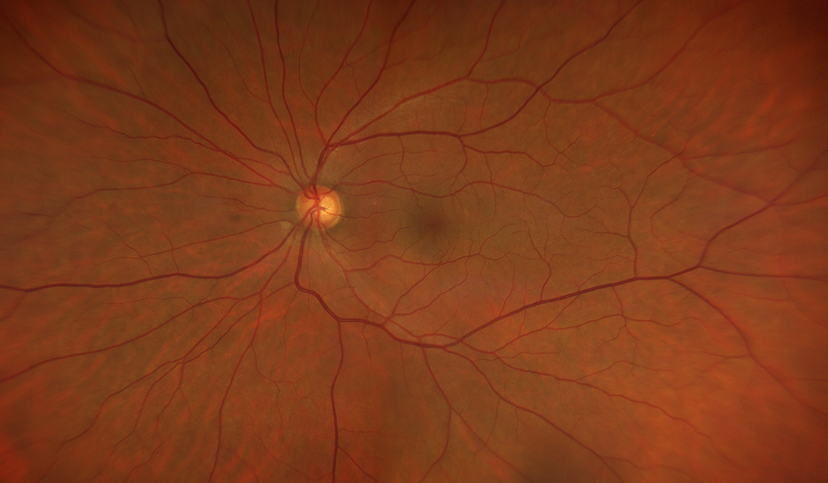
| Central light reflex affects the intensity profile of edge location, perhaps making a vessel appear thicker than it is. Photo: Ryan Schott, OD. Click image to enlarge. |
Studying retinal blood vessels can shed light on a number of conditions, including some beyond the eye. One property, vessel caliber, is thought to be a potential biomarker for pathologies such as atherosclerosis, coronary heart disease and some neurological diseases. However, researchers pointed out in a paper recently published in Translational Vision Science and Technology that there’s a lack of standardized assessment of retinal vessels, due in part to different modalities and technical factors, making clinical application challenging. They investigated potential bias from central light reflex and retinal vessel caliber agreement between two modalities, fundus camera and scanning laser ophthalmoscope, and reported that the two aren’t interchangeable.
The cross-sectional study included 85 eyes of 85 healthy individuals with different blood pressure status (aged 50 to 65). Patients underwent imaging with fundus camera and scanning laser ophthalmoscope, and the researchers measured central retinal artery equivalent (CRAE) and central retinal artery vein equivalent (CRVE).
They reported that the between-device difference in CRAE was greater than that in CRVE (9.5µm vs. 2.9µm). The fundus camera yielded higher measurements. The limits of agreement for the two modalities were -4.8µm to 23.9µm for CRAE and -12.0µm to 17.8µm for CRVE. The researchers noted that the between-device CRAE difference was positively associated with the presence of a central light reflex.
From a clinical perspective, the devices are “sufficiently interchangeable,” but the researchers noted that “nevertheless, color fundus images are likely to be more informative for assessing the central light reflex, whereas vessel diameter measurements derived from scanning laser ophthalmoscope images are less affected by the confounding effect of the central light reflex.”
“From an epidemiological perspective,” they added, “our results suggest that the effect of cardiovascular disease on retinal vessel caliber is likely to be underestimated, because the central light reflex makes arteriolar calibers appear wider in color fundus images.”
The researchers advocated for a standardized, objective protocol for retinal vessel caliber estimations. Given the bias from central light reflex, they recommended that future studies use vessel diameters adjusted for central light reflex for the most accurate estimates when assessing the relationship between retinal vessel caliber and ophthalmic or systemic disease.
Pappelis K, Jansonius NM. Retinal vessel caliber measurement bias in fundus images in the presence of the central light reflex. Transl Vis Sci Technol 2023;12:7:16. |
