  |
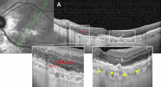
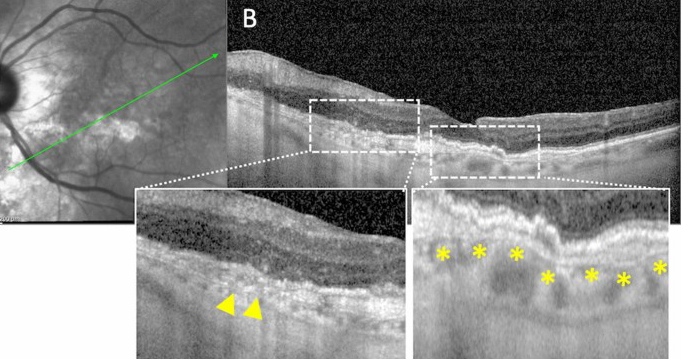
| While subclinical angioid streaks and AMD share some differences in pathogenesis, their shared features and high prevalence in the AMD population warrant consideration for angioid streaks as an AMD phenotype. The above images from the study show Bruch’s membrane dehiscences (a; red line), breaks (b; yellow triangles) and undulations (a, b; yellow asterisks). Photo: Sacconi R, et al. Ophthalmol Ther 2023. Click images to enlarge. |
The pathogenesis of age-related macular degeneration (AMD) isn’t completely understood, but there are several known phenotypes of the disease, each with unique pathogeneses and pathways. Studying these phenotypes may lead to targeted therapeutic approaches and new treatments, the thinking goes.
Recently, a team of researchers from Italy and the UK described what they consider to be a new AMD phenotype. Their findings, reported in Ophthalmology and Therapy, show that AMD patients with subclinical angioid streaks seen on OCT have different clinical presentation and imaging features, as well as disease pathogenesis, than AMD patients without this phenotype.
In the retrospective observational study, the researchers selected 274 AMD patients (543 eyes) who had no known causes for angioid streaks but whose eyes showed signs of that finding on structural OCT but not fundus examination. In particular, they examined Bruch’s membrane breaks and large dehiscences, which characterize angioid streaks.
A total of 73 eyes of 46 patients (mean age 81) showed angioid streak features on OCT that weren’t clinically visible. The researchers estimated the prevalence of subclinical age-related angioid streaks as 13.4% in this particular AMD population.
They reported that 73% of eyes with angioid streak features were affected by peripapillary atrophy, often with a petaloid pattern typical of angioid streak disease. A total of 97% of eyes presented with reticular pseudodrusen. Generalized equations showed that 41% of eyes with and 59% without drusen showed a significant difference in reticular pseudodrusen prevalence in angioid streak eyes on OCT vs. AMD eyes without subclinical angioid streaks.
Bruch’s membrane breaks were found in 100% of cases and dehiscences were found in 37% of cases.
“We suggest that subclinical angioid streaks should be considered to be a different phenotype in the AMD population, particularly with respect to possible new therapies,” the researchers wrote in their paper. “Indeed, since Bruch’s membrane likely plays a significant role in subclinical angioid streak etiology,” they speculated, therapeutic approaches can be different for this subtype.
“In particular, anti-complement treatments may not be as effective in this group of patients as in other drusen-driven ‘classic’ AMD phenotypes,” they continued. “An important future target is to identify which structure is predominantly involved in choroidal retinal aging, since this as yet unidentified structure could variably affect different anatomical sites.”
Sacconi R, Tombolini B, Zucchiatti I, et al. Subclinical angiod streaks with pseudodrusen: a new phenotype of age-related macular degeneration. Ophthalmol Ther 2023. [Epub August 5, 2023]. |
