 |
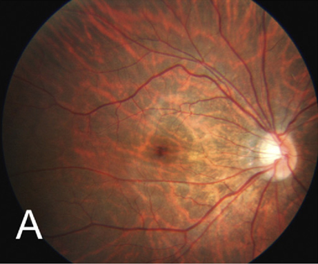
| In this study, 43.5% of highly myopic Chinese children had tessellated fundus (seen here in another patient) and 8.64% had diffuse chorioretinal atrophy. Photo: Hayashi K, Ohno-Matsui K, Shimada N, et al. Ophthalmology. 2010;117:1595-161. Click image to enlarge. |
Recognizing early signs of pathological changes in children with myopia can help to predict, diagnose and treat more severe disease later in life. One example is myopic maculopathy, which, despite its higher prevalence in adults, is still common among children. A recent study identified a progression rate of nearly 20% in young high myopes, highlighting the need for further studies on myopic maculopathy and associated choroidal and retinal changes in the pediatric population.
To help fill this gap in research, a team of investigators recently performed a cross-sectional study on 579 Chinese children aged four to 18 with high myopia (mean age: 12.8). Fundus photos were used to classify myopic maculopathy, while swept-source OCT was used to measure retinal thickness and choroidal thickness in the posterior pole.
The mean spherical equivalent of the cohort was -8.44D. The proportions of tessellated fundus and diffuse chorioretinal atrophy were 43.52% and 8.64%, respectively. Tessellated fundus was associated with a thinner macular choroidal thickness (odds ratio [OR]: 0.97), retinal thickness (OR: 0.98), longer axial length (OR: 1.55) and older age (OR: 1.13). The only factor that was independently associated with diffuse chorioretinal atrophy was a thinner macular choroidal thickness (OR: 0.94).
When using nasal macular choroidal thickness for classifying myopic maculopathy, the researchers found the optimal cut-off values to be 129.00µm for tessellated fundus and 83.85µm for diffuse chorioretinal atrophy.
“The results of this large-scale study of Chinese children with high myopia revealed that more than half could suffer from myopic maculopathy, in agreement with another recent study,” the researchers reported in their paper. “The choroid was significantly thinner, and the retina was only slightly thinner in children with more severe myopic maculopathy.”
One noteworthy finding that disagrees with previous research is the distribution pattern of choroidal thickness. In this study, “The thinnest peripapillary choroidal thickness was observed in the temporal sector in the groups with tessellated fundus and diffuse chorioretinal atrophy, suggesting a significant thinning of the choroid in the area between the macula and the optic disc,” the researchers explained. Conversely, in their last study, the team observed that “children with higher myopic degree tended to have a thinner peripapillary choroidal thickness only at the temporal and inferior sectors.” They suggest that the two studies’ contrasting results could be due to differences in the severity and course of myopia.
This data prompts concern regarding the high prevalence of children with myopic maculopathy. “The choroidal thickness in the posterior pole was closely associated with the severity of myopic maculopathy, and the nasal macular choroidal thickness can serve as a useful index for classifying myopic maculopathy,” the researchers concluded. They clarified that there is a need for further studies to confirm these findings.
Deng J, Xu X, Pan CW, et al. Myopic maculopathy among Chinese children with high myopia and its association with choroidal and retinal changes: the SCALE-HM study. Br J Ophthalmol. June 8, 2023. [Epub ahead of print]. |
