 |
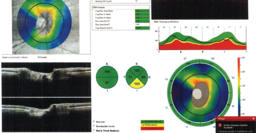
| Progressive cupping of the optic nerve head from retinal nerve fiber layer loss with corresponding visual field defects are characteristic of normal-tension glaucoma. Photo: Julia Reimold, OD, and Chris Wroten, OD. Click image to enlarge. |
Certain populations such as the Japanese have a high prevalence of normal-tension glaucoma, making risk factor identification critical to help catch these cases as soon as possible. Researchers recently investigated risk factors specific to the superior and inferior halves of the optic nerve head, since it’s thought that the process of glaucomatous damage in the two halves may differ.
They conducted a prospective study in which they followed 90 patients with NTG with untreated IOP (consistently ≤15mm Hg; mean IOP 12.3mm Hg) for five years, monitoring with Humphrey Perimeter and disc/retina stereo-photographs every three and six months, respectively.
They reported that the mean total deviation values significantly decreased at -0.5 and -0.13 dB/year in the superior and inferior hemi-VFs, respectively. In the superior hemi-VF, greater long-term IOP fluctuation was the risk factor for faster progression. In the inferior hemi-VF, disc hemorrhage, greater myopic refraction, body mass index and vertical cup-to-disc ratio were the risk factors for faster progression.
The researchers found that the progression probability was 47.7% and 17.7% at five years in the superior and inferior hemi-disc/retinas, respectively. Disc hemorrhage was a risk factor for both.
For some of these risk factors, such as long-term IOP fluctuation, the researchers pointed out that statistical detection sensitivity may vary between the superior and inferior half of the optic nerve head on both modalities due to lower progression rates in the superior half. “Therefore,” the researchers explained in their TVST paper, “the potential risk factors could be more sensitively detected in the inferior than in the superior half of the optic nerve head. In other words, the impacts of a higher body mass index, vertical cup-to-disc ratio and myopic power as a risk for further progression of glaucomatous damage should be more evident in the superior half of the optic nerve head than the inferior half of the optic nerve head.” Additionally, they noted that “the current result is not evidence that a higher long-term IOP fluctuation has no association with inferior hemi-VF progression.”
Asaoka R, Sakata R, Yoshitomi T, et al. Differences in factors associated with glaucoma progression with lower normal intraocular pressure in superior and inferior halves of the optic nerve head. Transl Vis Sci Technol 2023;12:8:19. |
