 A 38-year-old Hispanic female presented with decreased vision in both eyes that has persisted for the last four days. She experienced mild flu-like symptoms, a chronic headache and scalp tenderness approximately one week before the onset of blurred vision. Her ocular and medical histories were noncontributory. She takes no medications, and reported being otherwise healthy.
Upon examination, her best-corrected visual acuity measured 20/200 O.U. Extraocular motility testing was normal. Confrontation visual fields were full to careful finger counting O.U.
Amsler grid testing showed a large central area of metamorphopsia involving fixation O.U. Her pupils were equally round and reactive, with no afferent pupillary defect. Her intraocular pressure measured 15mm Hg O.U.
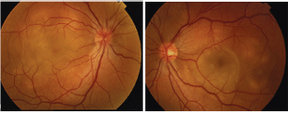
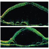
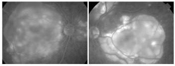
A dilated fundus exam revealed 1+ vitreous cell O.U. Her optic nerve evaluation showed small cups with good rim coloration and perfusion. There were significant changes in the maculae and posterior poles of both eyes (figures 1 and 2). Optical coherence tomography (OCT) (figures 3 and 4) and fluorescein angiography (FA) (figures 5 and 6) were also performed.
1, 2. Fundus images of our patient (O.D. left, O.S. right) show obvious retinal changes in both posterior maculae and posterior poles. 3, 4. Vertical OCT cuts of the maculae of both eyes (O.D. top, O.S. bottom). 5, 6. Late-phase fluorescein angiogram images of our patient (O.D. left, O.S. right) revealed these changes.
Take the Retina Quiz
1. What do the changes in the maculae and posterior poles of both eyes represent?
a. Multiple exudative retinal detachments.
b. Placoid lesions at the level of the retinal pigment epithelium (RPE).
c. Combined choroidal and retinal detachments.
d. Retinal pigment epithelitis.
2. What findings do the OCT and FA reveal?
a. Multiple retinal and RPE detachments.
b. Choroidal detachments.
c. Subretinal mass.
d. Hemorrhagic detachments of the RPE.
3. What is the correct diagnosis?
a. Sympathetic ophthalmia (SO).
b. Ocular sarcoidosis.
c. Vogt-Koyanagi-Harada syndrome (VKH).
d. Bilateral central serous retinopathy (CSR).
4. How should this patient be managed?
a. Observation.
b. Laser photocoagulation.
c. Intravitreal Avastin (bevacizumab, Genentech)
d. High-dose intravenous or oral steroids.
For answers, see below.
Discussion
The retinal changes seen throughout the maculae and posterior poles O.U. represent multiple exudative retinal detachments. The FA and OCT also demonstrate multiple RPE detachments.
Early in the FA, there was diffuse staining and leakage, mainly in the areas of the RPE detachments. As the angiogram progresses, you can see diffuse leakage of the fluorescein into the subsensory retinal space. In the later phases of the angiogram, you can truly see the extent and involvement of the detachments throughout the posterior pole.
The OCT not only demonstrates the bullous nature of the retinal detachment, but also the multiple small RPE detachments that are not visible on clinical examination.
So, what is wrong with our patient? The case history and clinical examination indicate that our patient has a fairly classic case of Vogt-Koyanagi-Harada syndrome.
The most significant finding in patients with VKH is bilateral granulomatous uveitis associated with exudative retinal detachments. Several other extraocular manifestations also may be presentmost commonly, pleocytosis of the cerebrospinal fluid.1 Other possible changes include vitiligo, poliosis, alopecia and dysacusis.1
The clinical course of VKH typically follows a pattern that begins with a prodromal stage, in which patients often develop a viral-like illness that lasts three to five days. During this time, patients may experience fever, malaise, headaches, orbital pain and nausea.1
Next, the disease transitions into the uveitic stage, in which patients develop blurred vision caused by exudative retinal detachments. Additionally, patients experience granulomatous inflammation such as thickening of the posterior choroid, edema of the optic nerve, and inflammation of the ciliary body and choroids.1 The inflammation may also include anterior chamber cell and flare.
Following the uveitic stage, the disease progresses to the chronic stage, in which patients develop depigmentation of the choroid. Depigmentation of the choroid associated with VKH has been referred to as the setting sun sign, in which the choroid and RPE take on a reddish hue. In this stage, patients may also develop vitiligio and poliosis.1
Finally, the chronic recurrent stage consists of a smoldering panuveitis, where patients can develop recurrent episodes of granulomatous anterior and posterior uveitis.1 The chances for recurrence largely depend on how early and adequately steroidal treatment is administered.
The diagnosis of VKH is typically based on clinical findings. There is some debate on the necessity of ancillary testing. In some less-straightforward cases, lumbar puncture and/or human leukocyte antigen (HLA) testing may be considered, depending upon the patients ethnicity.
VKH is most prevalent in people of Japanese ethnicity, accounting for 6.8% to 9.2% of all uveitic cases.2 The condition is also more common in individuals of Hispanic, American Indian or Asian Indian descent.1 HLA-DR4 testing has been shown to be very specific in detecting VKH in both Japanese and Korean patients. Interestingly, HLA-DR4 and HLA-DR1 testing accurately confirmed the presence of VKH in 89% of Mestizo patients in southern
Ultimately, proper and effective treatment governs the extent, severity and progression of VKH. Early and aggressive treatment with systemic corticosteroids is recommended, and the use of high-dose steroids may prevent progression and recurrence of the disease. The standard dosing regimen is 80mg to 100mg of prednisone q.d., with a very slow taper over six months. Or, intravenous methylprednisolone can be administered for three days, followed by a slow taper of oral steroids for up to six months.
Most VKH patients do very well on corticosteroid therapy and demonstrate a quick and rapid response. This includes complete resolution of the exudative retinal detachments and a return of the visual function.
We started our patient on oral prednisone at the standard dose. Within one week, her retinas reattached, and most of the fluid was reabsorbed.
1. Rao PK
2. Moorthy RS, Inomata H, Rao NA. Vogt-Koyanagi-Harada syndrome. Surv Ophthalmol 1995 Jan-Feb;39(4):265-92.
3. Arellanes-Garca L, Bautista N, Mora P, et al. HLA-DR is strongly associated with Vogt-Koyanagi-Harada disease in Mexican Mestizo patients. Ocul Immunol Inflamm 1998 Jun;6(2):93-100.
Retina Quiz Answers: 1) a; 2) a; 3) c; 4) d.
