 A 49-year-old Hispanic male presented to the emergency room with a chief complaint of a black spot in the vision of his left eye that he first noticed one day earlier. He was examined and reassured that there was nothing acutely wrong.
Two months later, the patient presented to our clinic for his annual eye exam. He reported that he still saw floaters in his left eye, but that his vision had remained stable.
On examination, his best-corrected visual acuity measured 20/20 O.D. and 20/30 O.S. Extraocular motility testing was normal. Confrontation visual fields were full to careful finger counting O.U. His pupils were equally round and reactive to light, with a trace afferent defect in his left eye. The anterior segment examination was unremarkable.
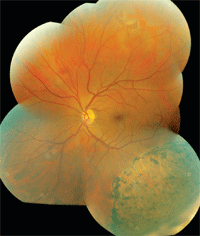
1. A fundus photomontage of our patient’s left eye. Note the retinal changes located inferiorly.
Fundus examination of the right eye showed a small cup, with good rim coloration and perfusion. The vessels, macula and periphery were normal. Examination of the left eye revealed a small cup as well as several obvious fundus changes (figure 1). A closer examination of the peripheral retina in the patient’s left eye can be seen in figure 2.
Take the Retina Quiz
1. How do you explain the changes in the inferior retina of the left eye?
a. Early retinitis pigmentosa (RP).
b. Congenital hypertrophy of the retinal pigment epithelium (CHRPE).
c. Previous retinal detachment.
d. Chronic retinal detachment.
2. What other significant retinal changes can be seen in the fundus photos?
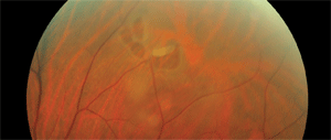
A closer view of our patient's left eye. What do you see?
a. Horseshoe retinal tear(s).
b. Retinal detachment.
c. Atrophic retinal holes.
d. Lattice degeneration.
3. How should this patient be managed?
a. Observation.
b. Pars plana vitrectomy (PPV) and a scleral buckle procedure (SBP).
c. Laser photocoagulation.
d. Gas fluid exchange and laser.
4. Why do you believe this patient was told that nothing was wrong with his left eye just two months before his scheduled eye examination?
a. The ER doctor who examined him likely missed the retinal findings.
b. The ER doctor did not believe that the findings were indicative of a significant problem.
c. The patient developed these fundus changes between the ER visit and the scheduled examination.
d. It is impossible to explain.
For answers, see below.
Discussion
Our patient has at least three horseshoe retinal tears––a large tear that is clearly visible in the fundus photograph, and two smaller tears next to it. Also, it’s important to note the bridging retinal vessel located in the larger tear—additional traction on the flap of the tear could cause the vessel to avulse, which could lead to a vitreous hemorrhage. Perhaps increased traction is already occurring, which would explain why our patient sees floaters.
The changes seen inferotemporally are also very interesting. Beyond the inferior arcade, you can see an arcuate pigmented demarcation line. Within that area, there is bony pigment spiculing and atrophic retina. Those findings tell a very interesting story. In the absence of any history, we know that this patient had a previous retinal detachment in this area. The pigment demarcation line is the boundary of the posterior border of the detachment. As the detachment resolved, the retina flattened and became scarred and atrophic, which allowed pigment to migrate from the RPE into the sensory retina.
Fortunately, we do have a history that extends beyond the patient’s ER visit. He originally presented nine years earlier with a five-day history of sudden visual acuity loss in his left eye. His visual acuity measured 3/200 O.S., and the clinician discovered a macula-off retinal detachment. The detachment included more than 180° of retina––extending from the 12 o’clock position down to 7 o’clock––with involvement of most of the temporal retina and macula. A retinal dialysis three clock hours in size was seen inferotemporally at the ora serrata and was identified as the cause of the retinal detachment. The patient underwent a successful pars plana vitrectomy, cryotherapy and a scleral buckle procedure. Amazingly, his acuity returned to 20/30, where it had remained since that time. This history explains the presence of the afferent pupillary defect and the 20/30 visual acuity.
Now, we need to try to answer three major questions:
• Does his history of retinal detachment nine years ago have anything to do with the horseshoe tears?
It’s difficult to know the answer to that question. Horseshoe tears most commonly develop as a result of traction at the posterior margin of the vitreous base. The vitreous base represents a circumferential zone where the vitreous is attached to the peripheral retina. It measures approximately 2mm to 6mm in width and straddles the ora serrata. The adhesion of the vitreous at the peripheral retina is evenly distributed circumferentially. Through the natural aging process, the vitreous begins to liquefy and irregularities develop in the shape and extent of the vitreous base. These irregularities result in isolated areas of focal traction that may promote the development of a horseshoe tear. These irregularities are likely responsible for the majority of retinal tears that are not associated with lattice degeneration.1
• Is increased traction the reason for our patient’s retinal tears?
This too is a difficult question to answer. Given that he already underwent vitrectomy years before, it seems unlikely that there would be any vitreous left to cause traction. It is possible that some residual vitreous remained from the vitrectomy years before and is now causing the traction. Or, perhaps the retina was weakened from a previous retinal detachment years before the surgery. While the patient’s history was not significant for trauma, the retinal dialysis indicated that the patient did in fact suffer trauma at some point. So perhaps some traumatic event many years earlier could explain all that has since happened.
• Finally, was the horseshoe tear indeed present at the patient’s ER visit?
Once again, there’s no way to know for sure. With the new onset of symptomatic floaters, it would suggest that something was occurring two months earlier. But, who knows if the area was even large enough to be seen on indirect ophthalmoscopy?
Thankfully, the patient’s retina did not detach during the two months before his eye examination. A retinal specialist saw the patient on the same day and immediately applied laser photocoagulation to seal off the retinal tears. The patient was seen a month later for follow-up and was doing well.
1. Wilkinson CP, Rice TA. Michels Retinal Detachment, 2nd ed. St. Louis: Mosby, 1997; 935-77.
Retina Quiz Answers: 1) c; 2) a; 3) c; 4) d.
