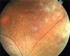
 A 31-year-old Hispanic male presented with sudden onset of blurred vision and photophobia O.S., and no prior ocular or systemic history. I noted significant cell and flare in the anterior chamber, early synechiae, hypopyon, and extensive vitritis and retinitis. What is the differential diagnosis?
The differential diagnosis for this patient falls into two broad categories: infection or inflammation, says Louis Pasquale, M.D., assistant professor of ophthalmology at
Apparently, the patient has not recently had surgery, which might be an avenue for infection, Dr. Pasquale says. Most likely, the patient has some inflammatory condition.
At first, his signs and symptoms might suggest an acute anterior uveitis. But, dont stop at the anterior chamber, says Anthony Cavallerano, O.D., of the VA Boston Health Care System. These patients often have anterior segment manifestations of posterior segment problems.
So, Dr. Cavallerano says, the differential diagnosis should encompass inflammatory conditions that also involve the posterior segment, such as ocular manifestations of sarcoidosis, syphilis, herpes zoster, tuberculosis, ankylosing spondylitis, endophthalmitis, or even Behets disease or acute retinal necrosis.
The next step: Take a thorough medical history. Id do a review of systems from top to bottom looking for clues for my next step, Dr. Pasquale says. For example, if this patient revealed he has HIV or AIDS, youd suspect cytomegalovirus retinitis.
Or, Dr. Cavallerano says, the patient might describe dermatologic or genitourinary symptoms that would alert you to Behet"s disease.
Depending on the information derived from the history, and assuming that the ophthalmic diagnosis is still unclear, consider ordering laboratory tests.
If the patient says hes been having night sweats and has a productive cough with sputum, then youll want to test for conditions such as tuberculosis or sarcoidosis. Youll want to get a sedimentation rate, plan a purified protein derivative (PPD) skin test and get a chest X-ray, Dr. Pasquale says.
Or, if the patient has joint or back pain, consider testing for ankylosing spondylitis by employing histocompatibility laboratory testing, such as an HLA-B27, and also get an X-ray of the spine.
In this patients case, though, the ocular sign of hypopyon is a big red flag, Dr. Pasquale says. Because a hypopyon is such an ominous sign of a vision-threatening disorder, you need to immediately refer this patient to a uveitis expert for an ongoing workup. To continue to manage this patient without a specialists opinion is not a good idea.
This patient was diagnosed with acute retinal necrosis (ARN). But, treatment was delayed because it was initially misdiagnosed as primary anterior uveitis. What is the proper treatment and prognosis for this patient?
ARN accounts for 5% to 6% of uveitis cases in the

The visual prognosis is poor for a patient with acute retinal necrosis.
Image courtesy: Anthony Cavallerano, O.D.
Its highly unusual that a retinitis results in a hypopyon, Dr. Pasquale says. But its not unprecedented.1
Perhaps this underscores the point. The key is not to stop at the anterior chamber, Dr. Cavallerano says, because weve all seen situations in which the patient was treated with a mydriatic/cycloplegic and a topical steroid without any consideration of the posterior pole or the peripheral retina as well.
1. Enzenauer RW, Bradshaw DJ, Waterhouse WJ. Acute retinal necrosis presenting with hypopyon. Ann Ophthalmol 2000 Jun;32(2):151-2.

