 |
|
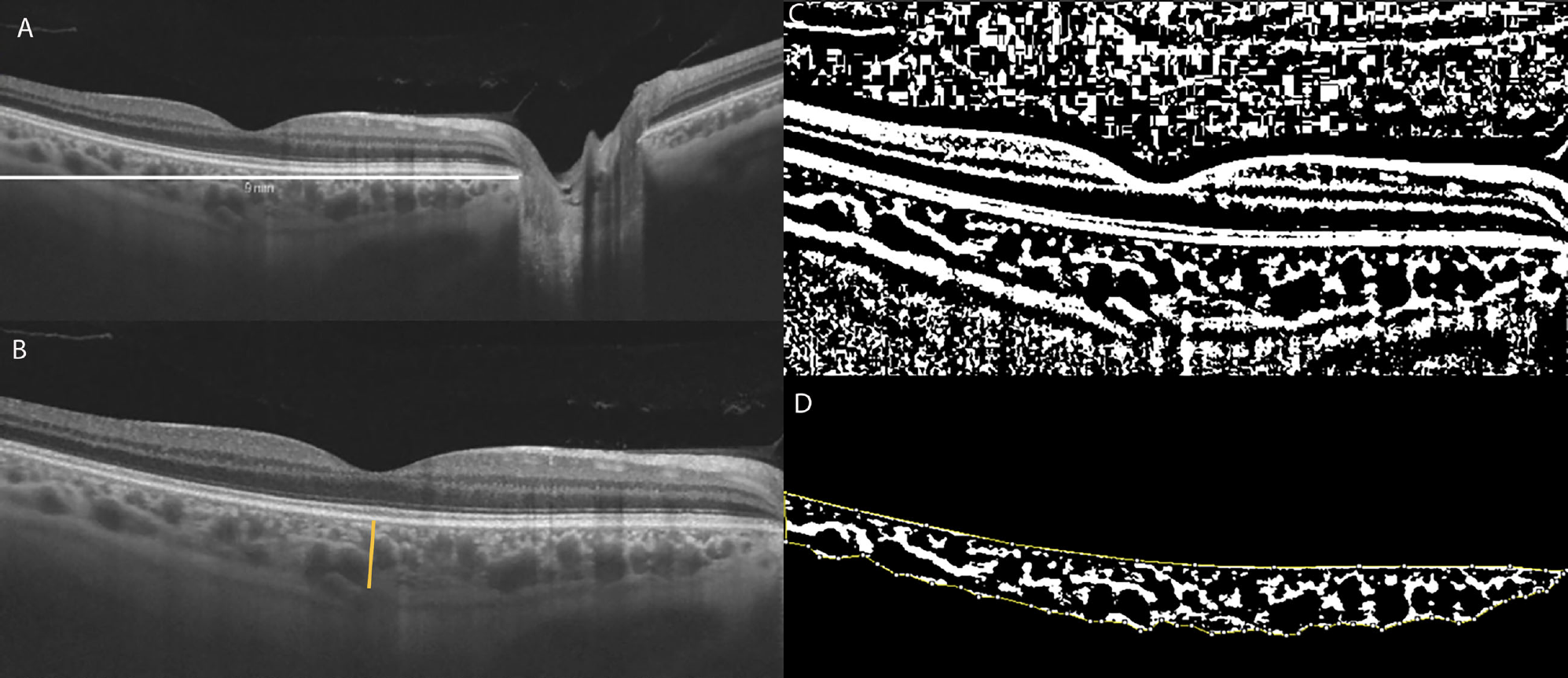
The CVI was the choroidal parameter most highly correlated with BCVA and might be a reliable imaging biomarker for monitoring progression. Dark pixels represent vessels’ luminal area and the white pixels the stromal area. Total luminal area divided by total choroidal area represents the CVI. Photo: Mucciolo DR, et al. Front. Ophthalmol. 2021; https://doi.org/10.3389/fopht.2021.755058.. Click image to enlarge. |
Prolonged or permanent visual impairment resulting from pathological myopia is among the more avoidable causes of vision problems worldwide, so it’s essential to investigate its pathogenesis and risk factors. Researchers recently evaluated changes in choroidal vasculature and their correlations with visual acuity (VA) in pathological myopia. They found that decreases in the choroid’s blood vessels were significantly correlated with visual impairment in myopia. Regarding axial elongation, the choroidal vascular index (CVI)—a newer OCT metric that estimates vascular capacity—decreased nonlinearly, whereas the other choroidal parameters decreased linearly. The CVI was the choroidal parameter most highly correlated with BCVA and might be a reliable imaging biomarker for monitoring the progression of pathological myopia.
The study cohort was comprised of 136 eyes from 80 participants, including 42 eyes from 21 participants with emmetropia/low myopia (-3.00D to +1.00 D), 48 eyes from 26 participants with simple high myopia and 46 eyes from 33 participants with pathological myopia. Eyes with spherical equivalent ≤-6.00D or axial length ≥26.5mm with diffuse or severe atrophy were classified as pathological myopia.
Swept-source OCT was used to image the eyes with a 12mm radial line-scan protocol. The parameters for 6mm diameters of the macula area centered on the fovea were analyzed. A custom deep learning algorithm was used to segment the choroidal boundaries. Then, the distance between the two boundaries was determined, and choroidal thickness, luminal area and stromal area were demarcated.
The CVI, choroidal thickness, luminal area and stromal area were lower in pathological myopia than in emmetropia/low myopia and simple high myopia. These values were also lower in simple high myopia than in emmetropia/low myopia, but there was no difference between the CVIs in the emmetropia/low myopia and high myopia groups.
Changes in CVI were not correlated with increases in axial length, and the study noted a critical flexion point in patients 40 and younger, approximately 27.26mm. “This indicates that the choroid might have been ‘overstretched’ in this stage, and the loss of choroidal vasculature was much more significant than that of the choroidal stroma in pathological myopia,” the researchers wrote in their paper for IOVS.
“Clinicians should pay close attention to alterations in the choroidal vasculature in pathological myopia and realize that preventing a decrease in the CVI might be a vital clinical goal for treatment,” they concluded.
Wang Y, Chen S, Lin J, et al. Vascular changes of the choroid and their correlations with visual acuity in pathological myopia. Invest Ophthalmol Vis Sci. 2022;63(12):20. |
