 |
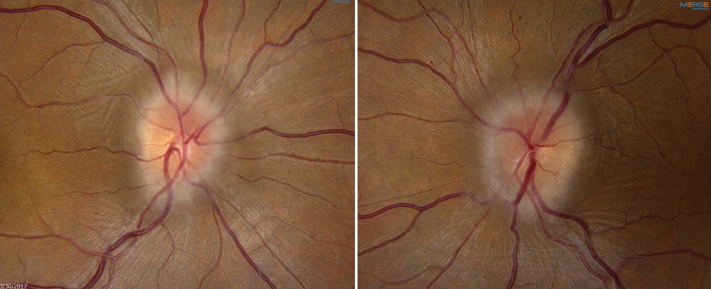
| Relapse triggers were present in most episodes of papilledema recurrence in the study cohort and included medication changes, weight gain and cerebrospinal fluid shunt malfunction. Photo: Mark Dunbar, OD. Click image to enlarge. |
Conventional wisdom has it that optic disc edema cannot occur in atrophic nerves, a supposition with consequences for the diagnosis and management of idiopathic intracranial hypertension (IIH). However, new research published in Journal of Neuro-Ophthalmology, demonstrated that optic discs with moderate to severe atrophy retain the ability to swell in response to raised intracranial pressure. Recurrences of atrophic papilledema were detectable on peripapillary OCT as changes in mean peripapillary RNFL thickness beyond the upper tolerance limit of test-retest variability. Peripapillary RNFL swelling in the absence of other features suggestive of relapse does not necessarily reflect a need for any treatment change, but it may indicate an opportunity for closer follow-up.
In a cohort of 165 patients with IIH, 32 eyes of 20 patients and 22 eyes of 12 patients demonstrated moderate and severe optic atrophy, respectively. Atrophy was defined as moderate if the average peripapillary RNFL thickness was ≤80μm and severe if the value was ≤60μm on at least two consecutive high-quality OCT scans. Based on the upper tolerance limit of test-retest variability, mean peripapillary RNFL elevation of ≥6μm with subsequent decrease to baseline thickness was considered papilledema.
Over a median follow-up of 198.5 weeks, 63.3% (19 of 30) of patients had at least one episode of relapse, and 50.0% (15 of 30) had at least one episode of papilledema. The rate, magnitude and concordance of peripapillary RNFL swelling were similar between the moderately and severely atrophic eyes. Final visual outcomes were similar between patients with and without papilledema recurrence.
There was a total of 36 relapse episodes, of which seven occurred in patients with clinical signs and symptoms but no OCT evidence of relapse, 12 occurred in patients with OCT changes but no clinical signs and symptoms of relapse and 17 occurred in patients with both clinical and OCT evidence to support relapse. Relapse triggers were present in most episodes of papilledema recurrence in the cohort and included medication changes, weight gain and cerebrospinal fluid shunt malfunction. Only 60.0% of patients with recurrent papilledema had worsening symptoms of raised intracranial pressure.
The median percent peripapillary RNFL increase in the latter two groups was 13.7%, and seven eyes (13.0%) of five patients (16.7%) showed thickening greater than 20.0% from baseline.
“In consideration of these factors, we suggest evaluating percent, rather than raw, changes in peripapillary RNFL thickness to monitor patients with idiopathic intracranial hypertension and concurrent optic atrophy,” the researchers wrote in their paper.
“Idiopathic intracranial hypertension relapse should be suspected if acute peripapillary RNFL elevation is bilateral, severe in magnitude, associated with a potential exacerbating factor and/or accompanied by worsening of symptoms, optic disc appearance on ophthalmoscopy or mean deviation on visual field testing,” they added. “OCT is helpful in monitoring patients with optic disc atrophy and idiopathic intracranial hypertension for treatment response and relapse, even when the degree of atrophy is severe.”
Xie JS, Donaldson L, Margolin EA. Swelling of atrophic optic discs in idiopathic intracranial hypertension. J Neuroophthalmol. July 12, 2023. [Epub ahead of print]. |
