 |
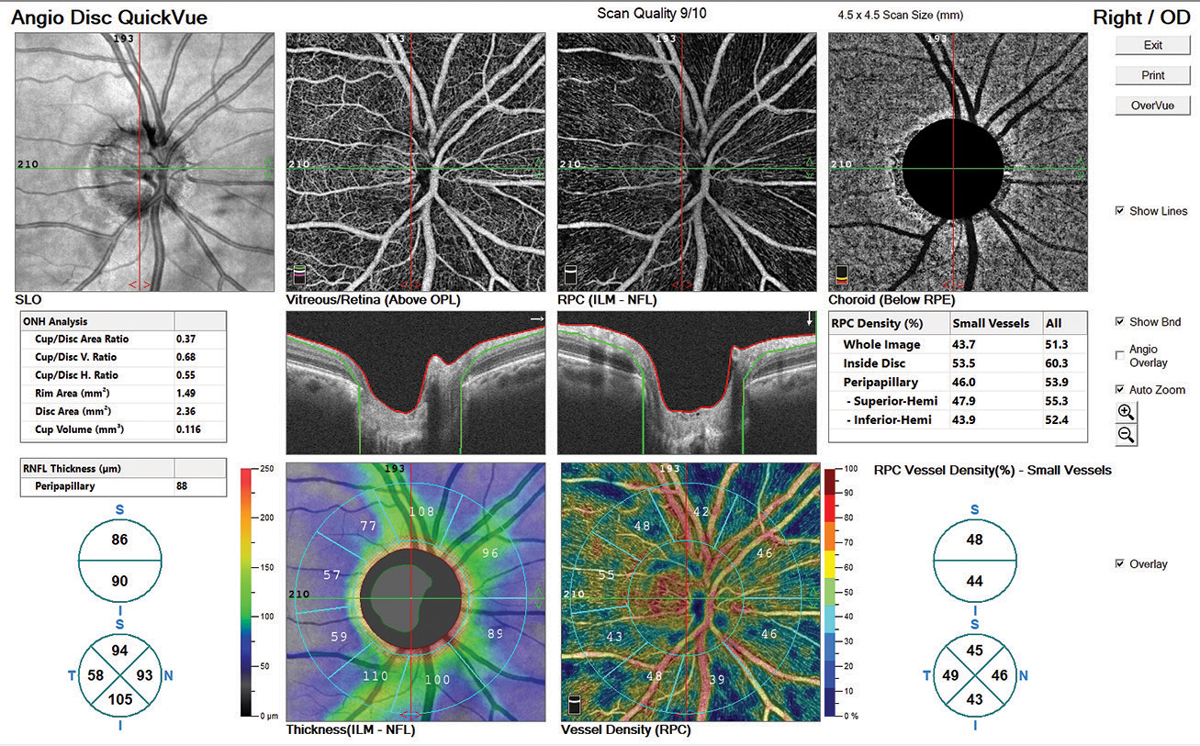
| For patients in the sample, vessel density was the only structural parameter to correlate with visual acuity. Photo: Ryan Schott, OD. Click image to enlarge. |
The assessment and management of glaucoma is important in all glaucoma patients, especially those with advanced and severe disease. At this stage, structural parameters may reach a point where no further loss is detectable. In a recent study, researchers aimed to evaluate the correlation between structural parameters and visual acuity (VA).
A total of 238 eyes were divided into an advanced glaucoma group and a severe glaucoma group. Structural parameters were obtained by RTVue optical coherence tomography angiography (OCT-A).
The deep nasal grid vessel density (VD) was the only structural parameter that was correlated with VA in patients with severe glaucoma. Compared with the superficial macular VD, which decreased in early glaucoma stages, the deep macular VD was compromised in later stages. The superficial macular VD showed better diagnostic accuracy in earlier stages.
“Among all grid VD, the VD of nasal grids had the highest area under the curve in both groups and retinal layers,” the authors explained. “It was the only OCT-A parameter that had correlation with VA in the severe glaucoma group. This area corresponded to the central papillomacular bundle, which was the last region to be damaged and maintained a good correlation with central visual function.”
Previous studies have reported that age and axial length affect VD. As this was also the case for this study, partial correlation was adjusted and a strong relationship between deep nasal grid VD and VA was observed in both groups.
This study agrees with previous ones investigating the relationship between VF sensitivity and OCT-A, “which had shown that VD had lower value for the measurement floor and was promising for disease monitoring in more advanced stages.
“In the advanced group, the superficial macular VD of different regions had excellent diagnostic accuracy; however, in the severe group, the deep macular VD of different regions and the foveal avascular zone density had acceptable discrimination.”
Patients with retinal diseases that may affect the macular VD were excluded, which means the decrease in deep macular VD could be solely attributed to glaucoma, the authors noted. They suggest future investigations focus on the longitudinal changes of macular VD and their association with functional progression.
Hsia Y, Wang TH, Huang Jy, et al. Relationship between macular microvasculature and visual acuity in advanced and severe glaucoma. Am J Ophthalmol. October 5, 2021. [Epub ahead of print]. |