 |
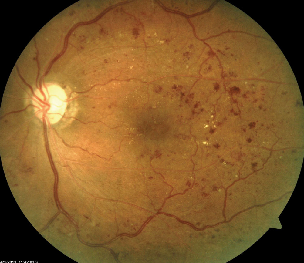
| Thinning in the inferior and superior quadrants of the pRNFL may help signify diabetic retinopathy. Click image to enlarge. |
A recent study determined a new clinical marker for identifying those with or at risk for diabetic retinopathy or diabetic retinal neurodegeneration. Structural changes such as a thinner superior and inferior peripapillary RNFL (pRNFL) were observed in those with higher glycated hemoglobin (HbA1c) levels and/or diabetes mellitus (DM), which researchers suggest may represent the area most affected by the disease.
Participants came from the EPIC-Norfolk Eye Study using data derived from the EPIC, a pan-European longitudinal prospective cohort study that began in 1989 to investigate lifestyle and environmental factors contributing to chronic disease. pRNFL measurements of each of the four quadrants (superior, inferior, nasal and temporal), as well as the average pRNFL thickness measures, were performed without pupil dilation using the GDxVCC. Of 8,623 participants who completed eye examinations between 2004 and 2011, 7,076 (12,555 eyes) had scan images of good enough quality to be included in the analysis.
All patients had HbA1c levels tested at the first (1993-1997), second (1998-2000) and third (2004-2011) study visit, and all three values were averaged to determine each participant’s estimated HbA1c. Patients considered to have DM needed to have either a self-reported history of DM diagnosis, use of DM medications and/or an average HbA1c ≥6.5%. If this information was absent from their records, the patient was only included in the HbA1c analysis rather than the one evaluating DM.
The researchers determined that for every unit increase in HbA1c, on average, inferior pRNFL thickness was 0.94µm lower, superior pRNFL thickness was 0.83µm lower and temporal pRNFL thickness was 1.33µm higher, each with a 95% confidence interval. The nasal pRNFL was the only quadrant that didn’t appear to be associated with HbA1c. When diabetes was used as the predictor rather than HbA1c, it demonstrated similar results.
“Our finding of lower RNFL thickness among individuals with high HbA1c levels (irrespective of DM status) is similar to studies in the neurology literature,” the researchers wrote in their paper. “In these studies, high HbA1c levels were associated with increased rates of central nervous system (CNS) neurodegeneration and decrease in memory score among older adults without DM. Proposed mechanisms underlying neuronal injury included increased formation of reactive oxygen species and advanced glycation end products in the setting of chronic hyperglycemia, which in turn promotes neuronal injury in the CNS. We believe that a similar neurodegenerative mechanism also occurs in the eye, that eventually manifests as RNFL thinning.”
The researchers also point out that some of the participants with high HbA1c levels may have had undiagnosed DM, as nearly half of those with the condition are unaware of it. They noted that the biomarker could have implications for the early detection of DM-associated neurodegeneration, perhaps before a patient even knows they have the condition.
“Recent studies have shown DM to be associated with abnormalities on brain MRI including regional reductions in brain volume in T1DM and global brain atrophy in T2DM,” they wrote. “MRI imaging, however, is more expensive and time-consuming and less easily accessible than optical coherence tomography (OCT). Hence, there may be a role for RNFL OCT imaging to serve as a potential biomarker for CNS volume loss in the future, if not for clinical, then for research purposes.”
The data from this study demonstrate that both diabetes and HbA1c levels are independently associated with thinner pRNFL in the superior and inferior quadrants, which researchers note should also be visible on SD-OCT, as its measurement accuracy has been shown to be comparable with the older device, GDxVCC, used in this study.
Zafar S, Staggers KA, Gao J, et al. Evaluation of retinal nerve fibre layer thickness as a possible measure of diabetic retinal neurodegeneration in the EPIC-Norfolk Eye Study. Br J Ophthalmol. December 24, 2021. [Epub ahead of print]. |