 |
|
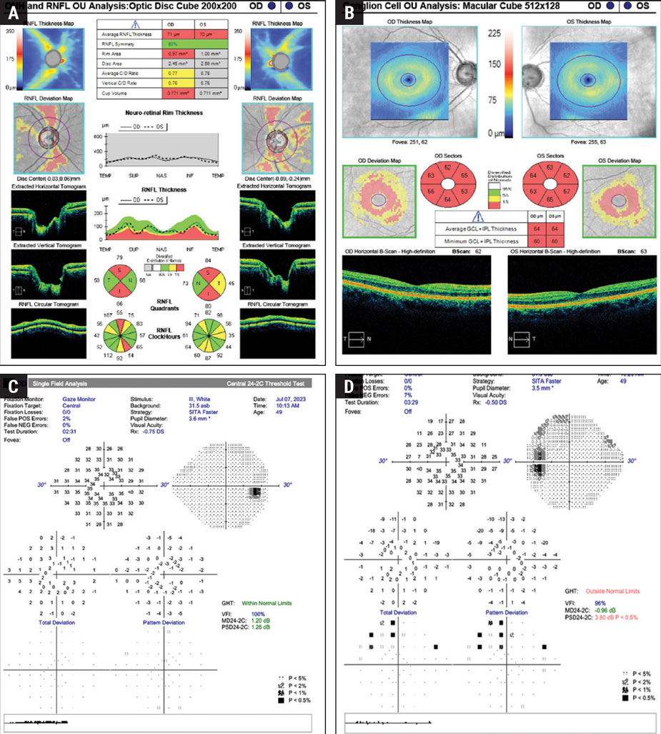
The 24-2C SITA Faster had good accuracy in detecting defects in mild-stage glaucoma, with 96.1% of patients found to have either central or beyond 10° VF defects. Photo: Brian Fisher, OD. Click image to enlarge. |
To detect mild-stage glaucoma with central visual field (VF) defects, the combination of the 24-2 SITA Standard and 10-2 SITA Standard is considered to be most accurate. Recently, a novel testing grid, the 24-2C, has been developed, which merged 10 additional points from the 10-2 test with the 24-2 testing grid, offering clinicians to focus on both central and peripheral vision in one test, or to opt for the 10-2 test for a more detailed central field assessment. Researchers in Japan aimed to determine the best program for detecting these defects in both the central and peripheral VFs in mild-stage glaucoma. The team examined patients, both with and without VF defects within the central 10°, with the 10-2 SITA Standard, 24-2 SITA Standard and 24-2C SITA Faster tests. They found the 24-2C SITA Faster test was highly effective and efficient for detecting mild-stage glaucoma with central VF defects and required markedly less time, which makes it a valuable tool in clinical settings.
This multicenter, retrospective cross-sectional study, which was published in American Journal of Ophthalmology, included 93 eyes with mild-stage glaucoma and central VF defects, along with 69 eyes with mild-stage glaucoma but without central VF defects. To compare the accuracy of the Center4, 24-2-12 and 24-2C-22 test points for detecting central VF defects in mild-stage glaucoma, receiver operating characteristic (ROC) curves were generated using logistic regression analysis after scoring four types of abnormal PD plots by using a sigmoid function.
The 24-2C test was significantly faster than both the 24-2 and 10-2 tests, reducing testing duration by 46% and 52%, respectively. In the upper-central VF, areas under the ROC curve of the total deviation (TD) plot were 0.50 for the Center4, 0.75 for 24-2-12, and 0.85 for 24-2C-22, with 24-2C-22 AUC significantly exceeding 24-2-12 AUC. For the pattern deviation (PD) plot, areas under the ROC curve were 0.53, 0.81 and 0.84, respectively. In the lower-central VF, using a total plot, areas under the ROC curve were 0.27, 0.57, and 0.57 for the Center4, 24-2-12 and 24-2C-22, respectively. Using the PD plot in the upper field, areas under the curve were 0.27, 0.64 and 0.81, respectively, with the area under the curve of the 24-2C-22 significantly exceeding that of the 24-2-12.
The analysis indicated that the prevalence of central VF defects in the lower field was higher on the temporal side, within 10°, than in the upper field. The additional points in the 24-2C test were placed more towards the temporal side, within 10° in the lower field than in the upper field. “These factors may explain the significantly higher area under the ROC curve for the 24-2C-22 test points when using the PD plot for central superior VF defects,” the study authors wrote in their paper.
“Since the patient group was limited to those with mild glaucoma, we considered that the PD plot reflects the actual sensitivity of the VF better,” they added. “It is possible that abnormalities within the central 10° could be detected more accurately with the 24-2-12 test points. This could explain the significantly higher AUC for the 24-2C-22 points than that for 24-2-12 points only when using the TD plot.”
The team also suggested that it is possible that the newest test’s lower detection accuracy in the lower field was related to the smaller number of patients with lower-field defects, as opposed to those with upper-field defects. The study concluded that the 24-2C SITA Faster test was highly effective and efficient for detecting mild-stage glaucoma with central visual-field defects. This, and its reduced duration, makes it a valuable tool in clinical settings.
Nishijima E, Fukai K, Sano K, et al. Comparative analysis of 24-2C, 24-2, and 10-2 visual field tests for detecting mild-stage glaucoma with central visual field defects. Am J Ophthalmol. July 31, 2024. [Epub ahead of print]. |