 |
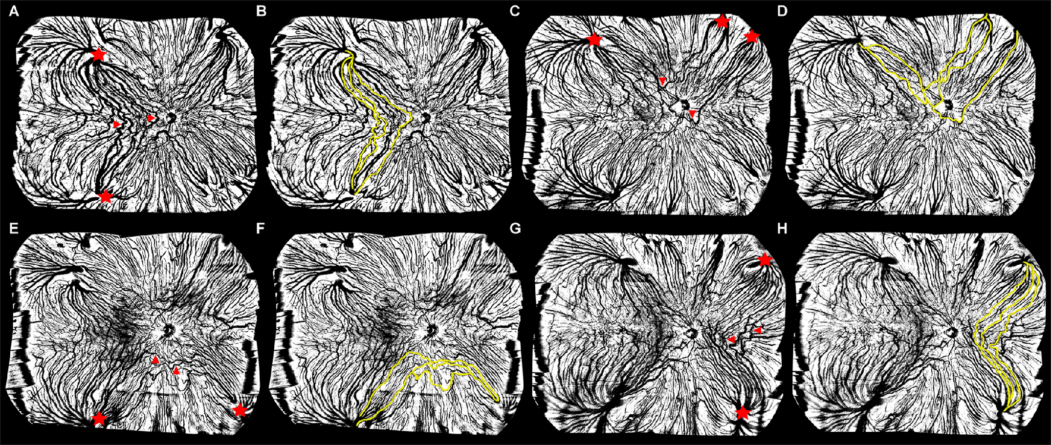
| The superotemporal quadrant of the vortex vein drainage system was found to have the thickest choroidal layer and highest choroidal vascularity index. This image from the study shows intervortex venous anastomoses in the temporal (A), superior (C), inferior (E) and nasal (G) quadrants, highlighted in yellow in images B, D, F, and H. Red stars indicate the vortex vein (VV) ampullas in adjacent quadrants, whereas the red arrows refer to the anastomotic vessels connecting the VV ampullas in adjacent quadrants. Photo: Luo Z, et al. Transl Vis Sci Technol. 2024;13(9):191. Click image to enlarge. |
The choroid’s vortex veins (VVs) are segmented into four distinct drainage systems that control choroidal venous outflow. Recent research has begun to focus on the function of this system in pachychoroid spectrum disorders using ultra-widefield OCT angiography (UWF-OCT-A); however, without a detailed understanding of the VV drainage system in healthy eyes, it’s challenging to interpret the findings of these studies. To help paint a clearer picture of the characteristics of this choroidal structure in healthy individuals, researchers recently performed an observational study on 207 eyes of 207 subjects in China.
UWF-OCT-A imaging was performed on all participants to measure choroidal thickness and vascularity index across different quadrants of the VV system. Comprehensive ocular examinations were also completed.
The results, published recently in Translational Vision Science & Technology, indicated significant variations in choroidal thickness and vascularity among different VV quadrants. The superotemporal quadrant typically showed the highest values, suggesting a preferential drainage system, which the researchers note has been shown in other studies that used indocyanine green angiography.
The study also revealed sex-related differences in choroidal vascularity, with men displaying higher indices than women. The researchers elaborated in their paper: “The choroid layer in the submacular area, as well as in the VV drainage quadrants, was found to be thicker in men than in women, although this difference was not significant.” Additionally, they noted that men attained a higher choroidal vascularity index than women not only in the submacular region, but all four VV drainage quadrants. Since CVI is considered a more stable biomarker of choroidal vessels than choroidal thickness, these results “support the observation that men experience higher perfusion in their choroidal VV circulation system than women.”
Choroidal perfusion in the VV drainage system also appeared to gradually decline with age, which the researchers suggest could be attributed to dysregulation of the sympathetic nervous system and vascular impairment.
Another notable finding was that almost half of healthy eyes had intervortex venous anastomoses (IVA) in the choroidal vessel networks, most commonly in the temporal quadrant (33.8% of eyes). Furthermore, the frequency of IVA in the macular region was 15.9%.
“The IVA in the temporal quadrant is also a common feature of pachychoroid spectrum disorders, which are characterized by increased choroidal thickness and vessel dilation,” the researchers wrote in their study. “As a result, we hypothesize that temporal IVA, especially macular IVA, may be considered a risk factor indicating a subclinical state of pachychoroid spectrum disorders.”
Some limitations of this study include the ethnic homogeneity of the sample (only Chinese participants) and UWF-OCT-A’s lack of ability to measure the actual blood flow velocity within the VVs. Additionally, indocyanine green angiography was not conducted to assess vessel permeability.
The authors concluded that based on the characteristics of the choroidal VV drainage system in healthy eyes, this choroidal structure “may be considered a promising biomarker for ocular diseases.”
| Click here for journal source. |
Luo Z, Xu Y, Xiong X, et al. Characterization of vortex vein drainage system in healthy individuals imaged by ultra-widefield optical coherence tomography angiography. Transl Vis Sci Technol. 2024;13(9):191. |