| |
|
| Vol. 2, #02 • Thursday, March 25, 2021 |
|
|
|
|
| |
|
Review's Chief Clinical Editor
Paul M. Karpecki, OD, FAAO
Provides you with cutting-edge clinical strategies for optimal management of ocular surface disease and beyond.
|
|
|
|
|
|
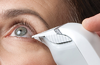 |
| Osmolarity testing |
|
If you’ve been working on a dry eye clinic for some time and want to advance or differentiate it, here are technologies you should consider.
The first is to adopt the use of tear osmolarity (TearLab). This technology has proven itself to be a reliable tool in my practice and highly accurate, according to research on diagnostic devices for dry eye1. Signs and symptoms don’t always align for dry eye patients or at least can be variable, so having an objective test is a necessity. Most other tests—including corneal staining, tear film breakup time, and even meibomian gland expression—are subjective.
With tear osmolarity, it’s key to have a well-trained technician who knows how to acquire tear samples consistently. Because the measurement occurs on the bottom (not the end) of the test card, proper alignment on the tear meniscus is essential for accuracy. Hitting the conjunctiva will result in reflex tearing, which will affect the measurement. It’s important to remind patients to avoid using any drops in their eyes for at least an hour before testing. A normal osmolarity measurement should be under 308 mOsmol/L, and stability is indicated by both eyes being within 6 mOsmol/L of each other. For example, a reading of 303 and 315 indicates moderate dry eye and instability, while a reading above 330 indicates severe dry eye. Normal measurements in a non-treated dry eye patient may point to a differential diagnosis such as epithelial basement membrane dystrophy (EBMD), allergic conjunctivitis, conjunctivochalasis, or eye misalignment, to name a few.
Another diagnostic tool for the advanced dry eye practice is meibography. In many patient cases, the meibomian glands will not yield any expression. This could be because a patient has few to no meibomian glands or has a complete set of very obstructed meibomian glands. Only meibography will help confirm this finding. The resulting management is analogous to glaucoma; the more severe the meibomian gland loss, the more aggressive one has to be. So it’s important to know the patient’s level of gland atrophy.
Another option is to purchase a comprehensive corneal diagnostic device such as the Keratograph M5 (Oculus), which includes a dry eye diagnostic suite of meibography, tear film breakup time, tear meniscus height measurement, and lipid layer evaluation, among other capabilities.
If you are serious about establishing an advanced dry eye clinic, these next-level diagnostic capabilities will prove extremely beneficial.
|
|
|
|
KEY TAKEAWAY: To evolve into an advanced dry eye disease practice, consider adding osmolarity and meibography capabilities.
|
|
|
|
| Supported by an independent medical grant from Kala Pharmaceuticals |
|
| |
| |
Review of Optometry® is published by the Review Group, a Division of Jobson Medical Information LLC (JMI), 19 Campus Boulevard, Newtown Square, PA 19073.
To subscribe to other JMI newsletters or to manage your subscription, click here.
To change your email address, reply to this email. Write "change of address" in the subject line. Make sure to provide us with your old and new address.
To ensure delivery, please be sure to add revoptom@lists.jobsonmail.com to your address book or safe senders list.
Click here if you do not want to receive future emails from Review of Optometry. |
|
|
|
|
|
|
|
|