With the escalating global prevalence of myopia, tilted optic disc in myopes has become increasingly recognized as a concern and garnered significant attention in recent years. Given the crucial role of retinal nerve fiber layer (RNFL) thickness in the diagnosis and prognosis of glaucoma, quantifying tilted optic disc could contribute to identifying the clinical significance of myopia in glaucoma pathogenesis, particularly in revealing potential mechanisms for heightened susceptibility and early assessment of glaucomatous optic neuropathy in myopia.
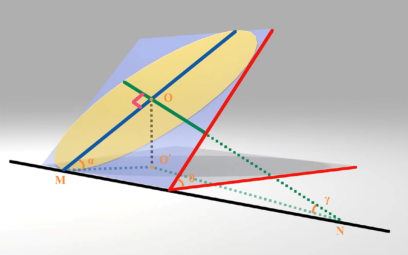 |
| A recent study revealed a more tilted optic disc tended to be associated with a longer AL. Considering the connection between deformations of the optic disc and peripapillary region and the scleral extension on the posterior eyeball segment during the axial growth of myopic eyes, this novel tilted optic disc measurement can contribute to further exploring potential pathologic changes caused by myopic-related axial extension. This image from the study shows a diagram of how discs were measured: the “ovality” of the disc (yellow oval) and the angle formed between the disc plane and coronal plane (red angle) were both measured and then correlated with RNFL values. The blue line represents Bruch’s membrane opening. Photo: Li Y, et al. Transl Vis Sci Technol. 2024;13(9):24. Click image to enlarge. |
A limitation of these established OCT-based approaches is that measuring the tilt angle of a tilted disc horizontally or vertically only indicates the tilt degree in a specific direction. By adopting a unique design principle that involves dividing the tilted optic disc oblique plane and constructing a mathematical model based on two distinct yet interconnected tilt angles measured through high-resolution cross-sectional SD-OCT images of the optic disc, researchers in Shenyang, China introduced a new three-dimensional method for objectively calculating disc tilt in the myopic population. They determined that a higher degree of myopia indicated a greater tilt angle of the tilted optic disc, and a greater tilted optic disc suggested additional changes in circumpapillary RNFL (cpRNFL) thickness. These findings should be considered when interpreting increased susceptibility and early assessment of glaucoma in myopia.
The study included the right eyes of 243 healthy young individuals, categorized by axial length (AL). The team measured the “ovality” of the disc’s shape as well as the “dihedral” angle (defined as the angle between the optic disc plane and the coronal plane) using SD-OCT infrared ray fundus photographs and high-resolution cross-sectional images of the optic disc, respectively. The relationships between these and other ocular-related parameters were analyzed.
Eyes in the longer axial length group exhibited a lower ovality index and a higher dihedral angle, along with a thinner nasal and inferonasal circumpapillary retinal nerve fiber layer (cpRNFL) and thicker temporal and superotemporal cpRNFL. There was a significant relationship between disc angle and cpRNFL thickness. The new method using dihedral angle to measure the tilt angle of the tilted optic disc demonstrated high repeatability.
“These methodologic innovations and enhancements enabled us to dimensionally transform tilted optic disc assessment, providing beneficial information to reflect the tilt degree of the optic disc plane relative to the coronal plane of the posterior pole of the eyeball,” the study authors wrote in their paper.
“The dihedral angle could be a superior indicator for evaluating the morphologic characteristics of myopic optic discs as AL increases, providing a basis for further investigations into myopic-related pathologic changes. In understanding abnormal RNFL thickness, the influence of tilted optic disc and its tilt degree should be recognized in the field of myopic glaucoma,” the study concluded.
| Click here for journal source. |
Li Y, Jia W, Liu X, et al. Measurement of the tilt angle of the optic disc using spectral-domain optical coherence tomography and related factors in myopia. Transl Vis Sci Technol. 2024;13(9):24. |