 |
|
|
|
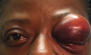
Gross exterior photographs of our 57-year-old patient who presented with visually significant facial and orbital injuries following a fall. What is the most likely diagnosis, and how should she be managed?
|
||
Diagnostic Data
Her best-corrected entering visual acuity measured 20/20 OD and 20/30 OS at distance and near, with no improvement upon pinhole testing. We observed no evidence of afferent pupillary defect OU. The biomicroscopic examination of the anterior segment revealed mild cell and flare OS. Intraocular pressure measured 15mm Hg OD and 20mmHg OS. There were peripheral pathologies OU. Our findings from the external evaluation are illustrated in the photographs.
Your Diagnosis
How would you approach this case? Does the patient require any additional tests? What is your diagnosis? How would you manage this patient? What is the likely prognosis?
Discussion
Additional evaluation includes ruling out a positive Seidel sign, iridodialysis, iridodenesis, peaked pupil (indicative of a penetrating injury), lens luxation, pupillary seclusion and hyphema. The diagnosis in this issue is a blowout fracture of the ethmoidal bones (medial wall) secondary to blunt trauma, with no evidence of muscle entrapment. Blunt trauma to the orbital rim is a frequent cause of orbital floor and medial orbital wall fractures.1-3 While there is no epidemiologic predilection for a blowout fracture, there are some clinical trends supported by national and regional databases.3,4
Patients who suffer blowout fractures are more likely to be male, range between the ages of 18 and 30, and engage in activities that include poor judgment either in or near the home.3,4 The term blowout fracture is reserved to describe an isolated orbital floor or medial wall fracture in the setting of an intact orbital rim.1-3 Patients who present with a history of blunt-force trauma often report being struck with a projectile (e.g., ball, bat or fist), suffering a collision injury (e.g., automotive airbag impact) or coming in contact with an object during a fall (e.g., staircase ledge).5
Following such an injury, pain, photophobia and lacrimation associated with post-traumatic uveal inflammation (iritis or iridocyclitis) are common.6 Other hallmark signs and symptoms include variable facial swelling secondary to fluid or air (orbital emphysema), crepitus (a crackling noise produced when air-infiltrated tissue is palpated), gaze-evoked diplopia, and pain upon eye movement.6 Associated collateral injuries include subconjunctival hemorrhage, ruptured globe, corneal abrasion, conjunctival laceration, hyphema, iridodialysis, lenticular subluxation, retinal detachment, vitreous hemorrhage, choroidal rupture and optic nerve evulsion.6 If the eye settles inferiorly or medially into the exposed sinus, enophthalmos with restricted ocular motility will be present, with or without loss of facial sensation. The seven bones of the orbit include the frontal, zygomatic, maxillary, ethmoid, sphenoid, lacrimal and pterygopalentine.7 By anatomic division, the roof includes the frontal bone’s orbital plate and the sphenoid’s lesser wing; the lateral wall is composed of the zygomatic bone and the greater wing of the sphenoid; the floor of the orbit is composed of the maxilla’s orbital plate, and the zygomatic and orbital process of the pterygopalentine bone; and the medial wall is composed of the maxilla, lacrimal and ethmoid bones, as well as the sphenoid body.7
The surrounding paranasal air sinuses comprise the other critical anatomical components.8 These include: medially, the ethmoidal air cells (anterior, middle and posterior); posterior and medially, the sphenoidal sinuses; inferiorly, the maxillary sinuses; and superiorly, the frontal sinuses.7,8 Several sinuses encircle the orbit, helping to lessen the weight of the skull and aid in voice resonance.8 Unfortunately, these structures exit the superior, medial and inferior walls of the orbit in an unsupported capacity, and are vulnerable to catastrophic failure via blunt-force trauma. There is some debate about the exact mechanism of a blowout fracture. When blunt force impacts the face, it may produce a combination of effects:9-12
• The force may strike the bone, produce a consequent shockwave, and cause “bone buckling.”
• The force may be transmitted by the globe, increasing orbital content pressure (i.e., a “hydraulic transfer”), yielding a fracture.
• The force may be transmitted to the eyeball, causing the globe to strike one of the orbital walls, resulting in a fracture. The point of breakage usually occurs along the axis of least support in an area where the tissue is the weakest.9-12
While all three aforementioned processes are described in the literature, bone buckling and hydraulic transfer are the most common mechanisms.10-12 Blowout fractures produced by the buckling mechanism are often limited to the anterior part of the orbital floor.11 In contrast, hydraulic fractures are often larger, involving both the anterior and posterior parts of the floor, as well as the medial wall of the orbit.11
In either case––because the orbital floor has been found to have a lower threshold for fracture than the medial wall and other orbital bones––when the orbital floor gives way, the globe and its attached components become unsupported. When this occurs, they slip down into the vacant sinus below, produce visible enophthalmos and gaze-evoked diplopia, as well as extraocular muscle dysfunction and infraorbital nerve hypoesthesia.10,12 For the optometrist, the treatment of blowout fractures centers around ocular first aid. Facial and orbital swelling literally can force the eye closed. The most challenging aspect of examining patients who have encountered facial blunt-force trauma is opening the eye for evaluation. In this instance, a Desmarres lid retractor can be used as a speculum to lift the superior lid.
Examination and management must proceed in a logical sequence from adnexa to posterior pole. This includes: imaging to rule out concomitant maxillofacial-orbital fracture or ruptured globe; treatment of facial lacerations with appropriate referral for cosmetic and functional closure; use of a Seidel test to rule out perforating injuries; application of topical anti-infective ointments for all cuts and abrasions; instillation of topical anti-infective agents for any observed corneal abrasion; and administration of topical and oral anti-inflammatory therapy for resultant ocular and facial swelling (i.e., topical cycloplegia, topical steroids, and topical and oral nonsteroidal preparations). Computed tomography remains the gold standard for assessing orbital fractures. In recent years, multi-slice CT technology has improved the acquisition of coronal orbit images without the need for neck hyperextension.13
Additionally, a dilated fundus evaluation is required to rule out vitreous hemorrhage, retinal tear and retinal detachment. Treatment of a blowout fracture may not be emergent. Clearly, if there is a compressive threat to the optic nerve via swelling and retrobulbar hemorrhage, the patient will require referral for an emergent lateral canthotomy and orbital decompression. Typically, surgical intervention is postponed until orbital health is consistent with a good surgical environment––unless large amounts of soft tissue are incarcerated in the bony rutpure.14
Orbital floor fractures traditionally have been managed via transconjunctival and subciliary incisions.15 However, postoperative lid malposition is a potential complication.15 Some surgeons have taken an endoscopic approach to manage orbital floor fractures, which features a hidden incision and improved fracture visualization.15 When the orbital floor requires replacement or reconstruction, ultra-thin, porous, polyethylene implants serve as durable substitutes that mimic the anatomy while avoiding the morbidity of rejection.16 We treated the patient’s left eye with 1% atropine BID and 1% prednisolone acetate Q2H, and instructed her to remain on bed rest. We advised her to use over-the-counter analgesics for additional pain control, as well as a nasal decongestant to avoid nose blowing. We also prescribed 500mg cephalexin PO BID as a prophylactic against infection. Over a two-week taper period, she improved without complication.
1. Lelli GJ Jr, Milite J, Maher E. Orbital floor fractures: evaluation, indications, approach and pearls from an ophthalmologist's perspective. Facial Plast Surg. 2007 Aug;23(3):190-9. 2. Burm JS, Chung CH, Oh SJ. Pure orbital blowout fracture: new concepts and importance of medial orbital blowout fracture. Plast Reconstr Surg. 1999 Jun;103(7):1839-49. 3. Guly CM, Guly HR, Bouamra O, et al. Ocular injuries in patients with major trauma. Emerg Med J. 2006 Dec;23(12):915-7. 4. McGwin G Jr, Owsley C. Incidence of emergency department-treated eye injury in the United States. Arch Ophthalmol. 2005 May;123(5):662-6. 5. Lehto KS, Sulander PO, Tervo TM. Do motor vehicle airbags increase risk of ocular injuries in adults? Ophthalmology. 2003 Jun;110(6):1082-8. 6. García-Medina JJ, García-Medina M, Pinazo-Durán MD. Severe orbitopalpebral emphysema after nose blowing requiring emergency decompression. Eur J Ophthalmol. 2006 Mar-Apr;16(2):339-42. 7. Snell RS, Lemp, MA. The orbital cavity. In: Snell RS, Lemp MA. Clinical anatomy of the eye, 2nd ed. 59-77. 8. Snell RS, Lemp, MA. The paranasal sinuses. In: Snell RS, Lemp MA. Clinical anatomy of the eye, 2nd ed. Malden, Mass.: Blackwell Science; 1998:78-89. 9. Burnstine MA. Clinical recommendations for repair of orbital facial fractures. Ophthalmology. 2002 Jul;109(7):1207-10. 10. Nagasao T, Miyamoto J, Nagasao M, et al. The effect of striking angle on the buckling mechanism in blowout fracture. Plast Reconstr Surg. 2006 Jun;117(7):2373-80. 11. Ahmad F, Kirkpatrick NA, Lyne J, et al. Buckling and hydraulic mechanisms in orbital blowout fractures: fact or fiction? J Craniofac Surg. 2006 May;17(3):438-41. 12. Rhee JS, Kilde J, Yoganadan N, Pintar F. Orbital blowout fractures: experimental evidence for the pure hydraulic theory. Arch Facial Plast Surg. 2002 Apr-Jun;4(2):98-101. 13. Cruz AA, Eichenberger GC. Epidemiology and management of orbital fractures. Curr Opin Ophthalmol. 2004 Oct;15(5):416-21. 14. Harris GJ. Orbital blow-out fractures: surgical timing and technique. Eye (Lond). 2006 Oct;20(10):1207-12. 15. Farwell DG, Strong EB. Endoscopic repair of orbital floor fractures. Facial Plast Surg Clin North Am. 2006 Feb;14(1):11-6. 16. Ozturk S, Sengezer M, Isik S, et al. Long-term outcomes of ultra-thin porous polyethylene implants used for reconstruction of orbital floor defects. J Craniofac Surg. 2005 Nov;16(6):973-7.
