 |
|
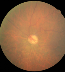
| Dilated fundus exam of our 77-year-old
patient's left eye. Her ocular history was significant for a central retinal vein occlusion OS. Now, we also suspect she has glaucoma. How should we proceed? |
Her best-corrected visual acuity measured 20/30 OD and light perception OS. Pupillary evaluation was significant for an afferent defect OS. Intraocular pressure measured 24mm Hg OD and 36mm Hg OS.
On gonioscopy, her angles were open OU. Slit-lamp evaluation revealed bilateral cataracts. Dilated fundus examination of the right eye revealed a cup-to-disc (C/D) ratio of 0.65 x 0.65, with a pink and distinct optic nerve. The left eye, however, exhibited a 1.0 x 1.0 C/D ratio and significant optic nerve cupping. Dilated fundus evaluation revealed no retinal or vascular changes OD, and small retinal hemorrhages, sclerotic retinal veins emanating from the optic nerve, and macular mottling OS.
Could undiagnosed glaucoma contribute to the CRVO in her left eye? Given the borderline IOP and moderate cup-to-disc ratio in her right eye, should we try to lower the pressure in order to prevent another retinal vein occlusion?
Glaucoma and RVO
The association between primary open-angle glaucoma (POAG) and RVO has been known for 100 years.1 POAG has been observed in more than 10% of patients who present with CRVO.1-4 It’s been stipulated that patients with a history of glaucoma may be up to five times more likely to develop RVO than those without glaucoma.2A few major case-control studies have reported a relationship between both conditions. A history of glaucoma or ocular hypertension (OHT) in the fellow eye is significantly more common in patients who develop CRVO compared to ocular non-hypertensive controls.5,6 Without question, age seems to be a primary confounding factor in this association.7-9
In the landmark Ocular Hypertension Treatment Study (OHTS), older patients who were diagnosed with OHT at baseline exhibited a statistically significant predictive risk for the development of RVO. While the average age of all OHTS participants was 55 years at the time of enrollment, those who developed RVO were more likely to be older than 65 years of age at baseline.7 Additionally, RVO occurred at an average of six years from baseline. It is important to note that data obtained from other trials, including the Beaver Dam Eye Study and the Blue Mountains Eye Study, further supported the relationship between glaucoma and RVO.8,9
Researchers have speculated that the relationship between POAG and RVO shares a similar pathogenesis to that observed between POAG and a disc hemorrhage.10 In eyes with concomitant POAG and retinal vein occlusion, the site of the RVO frequently is localized to the optic disc or located adjacent to the optic disc rim.10 There’s also a tendency for the occlusion site to occur at the area of the optic nerve head disc in patients who present with significant glaucomatous damage.
Finally, there’s been some debate as to whether POAG precedes or follows RVO development. One study indicated that, in most cases, the diagnosis of POAG took place prior to the presentation of the RVO.10 In patients diagnosed with POAG following the RVO presentation, there were obvious signs of glaucomatous disc damage and/or visual field defects prior to RVO development. These findings suggest that glaucoma typically is the precipitating ocular event in patients who present with both RVO and POAG.10
How Does POAG Cause RVO?
Due to the structural alterations at the lamina cribrosa induced by elevated IOP, distinguished ophthalmic researcher Frederick H. Verfoeff, MD, postulated that increased IOP likely was the leading risk factor for RVO development.1 He believed that increased IOP caused compression on the central retinal vein, which resulted in a CRVO.1 Additionally, retinal nerve fiber layer (RNFL) thinning has been regarded as a contributing factor to the development of a retinal vein occlusion. RNFL defects may cause a loss of structural support for a given retinal artery, causing it to collapse over the crossing vein, resulting in blood stasis. Blood stasis contributes to the development of thrombosis, which may cause RVO.4,11One study published in 2011 indicated that patients with RVOs typically have thinner RNFLs in the contralateral eye.12 This finding supports the possibility that glaucoma and RVO share a common vascular pathogenesis.12 Based on the literature, there seems to be a definitive causal relationship between POAG and RVO. Therefore, we decided to place our patient on Xalatan (latanoprost, Pfizer) QHS OD.
Because she was monocular and reported no associated pain, we elected not to treat her left eye. Additionally, we advised her to return in four weeks to assess the efficacy of treatment. Unfortunately, however, the patient was lost to follow-up.
Thanks to Marlon J. Demeritt, OD, of Oakland Park, Fla., for contributing this article.
1. Verhoeff FH. The effect of chronic glaucoma on the central retinal vessels. Arch Ophthalmol. 1913;42:145-52.2. Jeffrey S, Heier M, Morley G. Ophthalmology, vol. 8: Epidemiology and Pathogenesis of Venous Obstruction of Retina. Philadelphia: Mosby; 1999:18.3. Duane T. Clinical Ophthalmology. Revised, 3rd ed. New York: Harper and Row; 1981:2-3.4. Vannas S, Tarkkanen A. Retinal vein occlusion and glaucoma; tonographic study of the incidence of glaucoma and its prognostic significance. Br J Ophthalmol. 1960 Oct;44:583-9.5. The Eye Disease Case-Control Study Group. Risk factors for central vein occlusion. Arch Ophthalmol. 1996 May;114(5):545-54.6. Sperduto RD, Hiller R, Chew E, et al. Risk factors for hemiretinal vein occlusion: comparison with risk factors for central and branch retinal vein occlusion: the Eye Disease Case-Control Study. Ophthalmology. 1998 May;105(5):765-71.7. Kass MA, Heuer DK, Higginbotham EJ, et al. The Ocular Hypertension Treatment Study. A randomized trial determines that topical ocular hypotensive medication delays or prevents the onset of primary open-angle glaucoma. Arch Ophthalmol. 2002 Jun;120(6):714-20; discussion 829-30.8. Klein R, Klein BE, Moss SE, Meuer SM. The epidemiology of retinal vein occlusion: the Beaver Dam Eye Study. Trans Am Ophthalmol Soc. 2000;98:133-41; discussion 141-3.9. Cugati S, Wang JJ, Rochtchina E, Mitchell P. Ten-year incidence of retinal vein occlusion in an older population: the Blue Mountains Eye Study. Arch Ophthalmol. 2006 May;124(5):726-32.10. Lindblom B. Open angle glaucoma and non-central vein occlusion—the chicken or the egg? Acta Ophthalmol Scand. 1998 Jun;76(3):329-33.11. Hayreh SS, Zimmerman MB, Beri M, Podhajsky P. Intraocular pressure abnormalities associated with central and hemicentral retinal vein occlusion. Ophthalmology. 2004 Jan;111(1):133-41.12. Kim MJ, Woo, SJ, Park KH, Kim TW. Retinal nerve fiber layer thickness is decreased in the fellow eyes of patients with unilateral retinal vein occlusion. Ophthalmology. 2011 Apr;118(4):706-10.

