 |
History
A 55-year-old Cambodian female reported to the office with a chief complaint of decreased vision at near in both eyes. Her ocular history was unremarkable.
Her systemic history was remarkable for a lump in her neck, which was biopsied four years earlier.
She stopped going to the doctor when she became scared following the diagnosis of cancer.
She takes no medications and denies allergies of any kind.
Diagnostic Data
The patient’s best-corrected entering visual acuities were 20/20 in both eyes at distance and near through her PL/+2.50 bifocals. Refraction was stable with neglible differences.
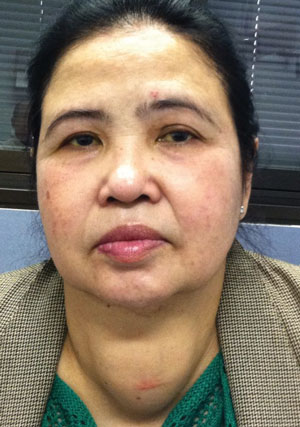
The pertinent external examination is demonstrated in the photograph.
The biomicroscopic examination of the anterior segment was normal. Goldmann applanation tonometry measured 13mm Hg in both eyes. The dilated fundus findings uncovered normal posterior poles, normal nerves and maculae with normal peripheral grounds.
Your Diagnosis
Does this case require any additional tests? What is your diagnosis? How would you manage this patient? What do you think is the likely prognosis?
Discussion
Additional studies included examination of old photographs, exophthalmometry, corneal sensitivity and photodocumentation of the external findings.
 | |
| This 55-year-old patient has a cancer diagnosis, but no remarkable ocular history. Now, she’s presenting with decreased vision at near in both eyes. Can you offer a diagnosis? |
This patient was diagnosed with a presumed thyroid goiter. The definition of a goiter is a thyroid gland that is larger than normal for a patient’s age and sex; specifically a volume of 25mL for men and 18mL for women.1 A goiter can be associated with hyperthyroid, hypothyroid and euthyroid (normal thyroid function) conditions, although in most of the world, the most common cause of goiter is iodine deficiency.1-3
In a normal functioning thyroid gland, iodine is needed to produce the thyroid hormones T3 (triiodothyronine) and T4 (thyroxine). If there is not enough iodine, the thyroid gland undergoes hyperplasia (increases in size), as its tissues attempt to manufacture the correct and necessary levels of hormones in the absence of the correct building blocks. The result is a goiter and as the thyroid organ fails under the stress, a hypothyroid state.2
In the United States, iodine deficiency is rare due to the ionization of salt. When goiters in the United States occur they are more likely to develop with or from other thyroid conditions.3 Hyperthyroidism is a type of thyrotoxicosis that causes excessive production of circulating thyroid hormones.
The most common cause of hyperthyroidism is Grave’s disease.2,4,5 Grave’s disease is an autoimmune disorder where circulating antibodies mimic the action of the pituitary-released thyroid-stimulating hormone (TSH). These antibodies bind and activate the TSH receptor on the thyroid, causing increased synthesis and release of thyroid hormones.2,4,5 The over-stimulation of the gland causes hypertrophy of thyroid follicular cells, resulting in a goiter.2 In addition to the goiter, the most common extrathyroidal feature of Grave’s disease is ophthalmopathy, which is present in roughly 50% of affected patients.5-7 The ophthalmic findings of Grave’s disease include bilateral, non-pulsatile proptosis due to extraocular muscle enlargement, impaired ocular motility, eyelid retraction, resistance to globe retropulsion, diplopia, decreased visual acuity, corneal exposure-related keratopathy and in the end stages, compressive optic neuropathy.5-8 Other conditions that can cause thyrotoxicosis include toxic nodular goiter, thyroiditis and exogenous ingestion of thyroid hormones.4
Hashimoto’s Thyroiditis is another potential cause of goiter formation.1,2, 9,10 In this autoimmune condition, white blood cells and antibodies, attack and invade the thyroid gland. In the initial stage of the disease, the thyroid gland can be overactive, underactive or normal, but over time hypothyroidism always develops.1,2,9 Hashimoto’s thyroiditis can be associated with other endocrine disorders, such as diabetes mellitus, Addison’s disease, hypo-gonadism, hypoparathyroidism as well as other autoimmune conditions, such as pernicious anemia, systemic lupus erythematosus and Sjogren’s syndrome.2,9
Thyroid cancer must always be ruled out when a goiter is present. Thyroid cancer accounts for 4.0% to 6.5% of all thyroid nodules.11 Thyroid cancer typically presents as nodules rather than diffuse thyroid enlargement.2 Although most thyroid nodules are benign, factors that increase the risk of malignancy include male gender, history of head or neck irradiation and either youth or advanced age. Nodules that are “hot” (producing excess thyroid hormones) are generally not malignant.10-11
Most patients with goiter have few or no symptoms.1-3,10,11 Thyroid enlargement to 40mL or more is generally visible, with patients presenting to their doctor for cosmetic reasons alone.1 Depending on the rate of growth and location, patients may exhibit symptoms that include dysphagia (difficulty swallowing) in retrosternal cases and dyspnea (difficulty breathing) in retrotrachael cases.1-3,10,11 Depending on the cause of the goiter, symptoms of hyperthyroidism or hypothyroidism may be present as well.2
TSH and thyroid hormone assays are the diagnostic tests of choice for patient’s presenting with a goiter. Low TSH levels suggest hyperthyroidism, while a high TSH levels suggest Hashimoto’s thyroiditis.1,3,4,10,11 Recent studies have suggested that TSH levels and serum thyroglobulin antibodies are also independent predictors of malignancy in patients with thyroid nodules.12,13 In addition to TSH and thyroid hormones, calcitonin should also be measured to rule out medullary thyroid carcinoma.11,14
Thyroid scintigraphy should be performed in cases where TSH levels are measured as low.1,10,11 This technique images the iodine-trapping ability in the nodule compared to the surrounding tissue.10,11 Since the test has low diagnostic value, it is only utilized to confirm the functional status of a suspected hyperfunctioning nodule.11 If the nodule is considered ‘hot’ it seldomly has malignant potential.10,11 Neck palpitation is a diagnostic tool used for detection and estimation of size and number of thyroid nodules. Ultrasonography has proven a more precise and sensitive test. If TSH levels are high or normal, ultrasonography should always be performed to confirm the presence of a mass, assess if the lesion is single or multiple and guide fine needle aspiration (FNA) biopsy.3,10,15 Up to 50% of patients with a single palpable thyroid nodule will demonstrate additional nodules on ultrasonography.10 Certain nodular features on ultrasonography are associated with increased risk of malignancy; nodule hypoechogenicity, the presence of microcalcifications, increased vascular flow, irregular borders and the absence of a halo.1,10
Thyroid FNA biopsy is the most valuable, cost-effective and accurate method in the evaluation of thyroid goiter.10,11,15,16 The goals of FNA biopsy are to detect malignant tumors and to prevent unnecessary surgery.1 Studies show that ultrasound-guided FNA biopsy is more specific and sensitive than palpation-guided FNA biopsy and should be considered the procedure of choice.15,16 Ultrasonography also assists in choosing the best targets for biopsy It is also helpful when the nodules are small, cystic and nonpalpbale.10,11
Treatment
The treatment of patients with goiters should be tailored to each patient. If the goiter is from iodine deficiency, correction with iodine supplementation should be the main objective.1 A combination of iodine with levothyroxine can also be given. The LISA study in Germany demonstrated that combination therapy of TSH-adapted levothyroxine with iodine was superior to levothyroxine monotherapy and iodine monotherapy alone at reducing the thyroid volume.17 This drug combination decreased the nodule size by 21.6%.17 It is important to note levothyroxine monotherapy is considered controversial, and should be avoided since it has the potential to cause subclinical hyperthyroidism, regrowth of the goiter and low bone mass in postmenopausal women.1,10 Drug therapy is cost-effective and non-invasive, but the lack of data on long-term success, iatrogenic hyperthyroidism, decreased compliance and ineffectiveness for large nodular goiters make it less successful than other treatment methods.1
Radioactive iodine therapy can reduce the volume of goiters by 35%-40% in one year and 40%-60% in two years. It is considered superior to drug treatment for reduction in thyroid volume.1,3,4
Goiters rarely return after therapy. When they do, the treatment can easily be repeated. The success in thyroid reduction depends on the age of the patient, duration of the goiter, size of the goiter, treatment activity level and homogenecity of iodine storage.1
One complication of the treatment is that it commonly results in hypothyroidism due to destruction of the gland.1,10,18 One study showed that of 265 patients, 32% had hypothyroidism at three months, 55% at one year and 73% at up to eight years of follow-up.18 There is no evidence of increased risk of thyroid cancer or other solid tumors after radioiodine therapy in adults, but no large studies have been done in children, therefore, it is not recommended before the age of 18-20 years.1
Thyroid surgery is indicated in multiple scenarios including; when cancer is suspected, very large or obstructive goiters and when there is restrosternal or medistinal extension of the goiter.1-3,10,11 The decision regarding how much thyroid tissue to resect is tailored to each individual case.1,3 Post-operative complications are uncommon, but do occur, including; recurrent laryngeal nerve palsy (permanent in 0.7% and transient in 2.9%) and postoperative hypoparathyroidism (0.5-6.6%).1,3,10 Hypothyroidism always develops after a subtotal thyroidectomy. Here, supplement therapy with levothyroxine is mandatory.3
In Grave’s disease, long-term treatment with anti-thyroid drugs, including methimazole and propylthiouracil, is the treatment of choice for patients with small goiters without ophthalmopathy.5 Drug therapy is not indicated in large goiters due to the likelihood of recurrent hyperthyroidism after drug withdrawal. If ophthalmopathy is present, radioactive iodine therapy or thyroidectomy is preferred.5
Management
If the patient is treated conservatively with either observation only or drug-therapy, follow-up examinations should occur every six to 18 months to assess thyroid gland volume changes, nodule changes in size or shape, formation of new nodules and change in thyroid function.1,11
Patients who have undergone radioactive iodine therapy or thyroid surgery require life-long follow-up due to the possibility of developing hypothyroidism.1,3 If hypoparathyroidism develops after thyroid surgery and lasts longer than six months, drug supplementation is necessary. Calcitrol and calcium are commonly the drugs of choice for this iatrogenic condition.1,11 If the cause of the goiter is thyroid cancer, further treatment is indicated.11
Finally, if Grave’s disease and ophthalmopathy are present, systemic and ocular treatment is indicated.5-8 Multiple studies are now investigating the use of oral Selenium in the mild stages of the disease, since serum selenoprotein P, an antioxidant, is commonly reduced in Grave’s disease patients.6,19,20 Selenium has shown to slow the progression of the disease, improve the quality of life and reduce resultant ocular disease.19 If the ophthalmopathy is moderate to severe, oral and intravenous steroids are usually the first line of treatment.5-8 If corticosteroid therapy fails or is not tolerated by the patient, biologics and monoclonal antibodies against TNF-α have demonstrated an ability to decrease inflammation and proptosis.6-8 If the ophthalmopathy is sight-threatening, orbital decompression is an effective treatment with lasting results.5-8
Goiters can be a complex clinical manifestation with many possible causes. It is important to determine the exact cause of the goiter and/or nodule with TSH testing, scintigraphy, ultrasonography and FNA biopsy so that treatment can be tailored to each individual patient.
Thyroid cancer should always be considered in every case and must be ruled out with testing. In addition to decreasing the thyroid volume itself, other treatment may be warranted depending on the etiology of the goiter.
Dr. Gurwood thanks Kelsey T. Moody, OD, for her contributions to this case.
1. Fuhrer D, Bockisch A, Scmid KW. Euthyroid goiter with and without nodules—diagnosis and treatment. Deutsches Arzteblatt International. 2012;109(29-30):506-16.2. Hershman J. Thyroid gland disorders. In: The Merck Manual Home Health Handbook. Whitehouse Station, NJ.: Merck Sharp & Bohme Corp; 2012.
3. Medeiros-Neto G, Camargo RY, Tomimori EK. Approach to and treatment of goiters. Med Clin N Am. 2012;96(2):251-368.
4. Sharma M, Aronow W, Patel L, et al. Hyperthyroidism. Med Sci Monit. 2011;17(4):RA85-91.
5. Menconi F, Marcocci G, Marino M. Diagnosis and classification of Graves Disease. Autoimmunity Reviews. 2014;13;298-402.
6. Stan MN, Garrity JA, Bahn RS. The evaluation and treatment of Graves ophthalmopathy. Med Clin North Am. 2012;96(2):311-28.
7. Sowka JW, Gurwood AS, Kabat AG. The Handbook of Ocular Disease Management, 11th ed. Review of Optometry. 2009;146(4):1-66.
8. Verity DH, Rose GE. Acute thyroid eye disease (TED): Principles of medical and surgical management. Eye. 2013;27:308-19.
9. Akamizu T, Amino N, DeGroot L. Hashimoto’s Thyroiditis. In: Thyroid Disease Manager. South Datmouth, MA: Endocrine Education; 2013.
10. Bahn RS, Castro MR. Approach to the patient with nontoxic multinodular goiter. J Clin Endocrinol Metab. 2001;96(5):1202-12.
11. Ross DS. Diagnostic approach to and treatment of thyroid nodules. UpToDate. Stanford Univ Sch of Med. 2013;1-21.
12. Haymart MR, Repplinger DJ, Leverson GE, et al. Higher serum, thyroid stimulating hormone level in thyroid nodule patients is associated with greater risks of differentiated thyroid cancer and advanced tumor stage. J Clin Endocrinol Metab. 2008;93:809-14.
13. Kim ES, Lim DJ, Back KH, et al. Thyroglobulin antibody is associated with increased cancer risk in thyroid nodules. 2010;20:885-91.
14. Nose V. Familial thyroid cancer: A review. Modern Pathology. 2011;24:S19-S33.
15. Can AS, Peker K. Comparison of palpation-versus ultrasound-guided fine-needle aspiration biopsies in the evaluation of thyroid nodules. BMC Reasearch Notes. 2008;1(12):1-5.
16. Kountakis SE, Skoulas IG, Maillard AA. The radiologic work-up in thyroid surgery: fine-needle biopsy versus scintigraphy and ultrasound. Ear, Nose & Throat Journal. 2002;81(3):151-4.
17. Grussendorf M, Reiners C, Pasche R, et al. Reduction of thyroid nodule volume by levothyroxine and iodine alone an in combination: A randomized, placebo-controlled trial. J Clin Endocrinol Metab. 2011;96:2787-96.
18. Kahraman D, Keller C, Schneider C. Development of hypothyroidism during long-term follow-up of patients with toxic nodular goiter after radioiodine therapy. Clin Endocrinol (Oxf). 2012 Feb;76(2):297-303.
19. Marcocci C, Kahaly GJ, Krassas G, et al. Selenium and the course of mild Graves’ Orbitopathy. N Engl J Med. 2011;364:1920-31.
20. Wetenbruch T, Willenberg HS, Cornelia S, et al. Serum selenium levels in patients with remission and relapse of Graves’ disease. Medicinal Chemistry. 2007;3(3):281-4.
