Control of a Pthirus pubis (crab louse) infestation of the eyelid involves mechanical removal of both the organisms and their egg cases or nits. The nits are often so firmly fastened to the lashes that they must be cut or epilated, which is irritating to both the patient and clinician. When a case presented in my office, I tried a slightly different approach that made treatment less painful for all involved.
History
A 74-year-old Hispanic female presented to our clinic complaining of slightly reduced distance vision and itchy eyes. Her medical history was significant only for a father with arthritisthe patient reported that she also had mild arthritis, for which she occasionally took Tylenol (acetaminophen, McNeil-PPC). Her ocular history was unremarkable.
She had been using Murine OTC eye drops (polyvinyl alcohol 0.5%/ povidone 0.6%, Prestige Brands) b.i.d. during the previous two to three weeks for the itching and mild irritation. She misplaced her glasses three to six months ago and presented without them.
Diagnostic Data
Her uncorrected visual acuity was 20/25 at distance and 20/50 at near O.D., and 20/40-2 at distance and 20/50-2 at near O.S. Best visual acuity improved to 20/20-2 at distance and 20/30 at near O.D., with a manifest refraction of
Pupils were equally round and reactive to light with a diameter of 1.5mm. Confrontation fields were normal O.U. Extraocular muscles were unrestricted in all gazes. Cover testing revealed orthophoria at distance and two to four prism diopters of exophoria at 40cm.
Anterior segment slit lamp examination revealed several crab lice attached to the lower lashes and feeding near the lower lid margins O.U. The lashes of the upper lid were thick with nit cases, but the lashes of the lower lid held few nits. The lower lid margins were crusty in appearance. The bulbar and palpebral conjunctivas were mildly injected. There were small pinguecula nasally O.U.
The patients corneas were intact and clear. The anterior chamber showed no cells or flare. The anterior chamber was open; the chamber angles were 4/4 by Von Herricks method. There was a grade 1+ nuclear sclerosis O.D. and a grade 2+ nuclear sclerosis O.S. of the crystalline lens. Intraocular pressure measured 15mm Hg O.U.
We deferred the dilated fundus exam to the follow-up visit, so that we could begin treatment of the lice infestation immediately.
Diagnosis
This patient had a Pthirus pubis infestation of the eyelids.
Treatment and Follow-Up
The patient and a family member who accompanied her were advised of the exam findings. As the family member looked on, a thick layer of bland ophthalmic ointment, Refresh P.M. (white petrolatum 57.3%/mineral oil 42.5%, Allergan) was applied to both the upper and lower lids. I advised the patients relative to renew this application in the same way q.i.d in order to assure complete and uninterrupted coverage.
The probability of infestation in other areas of the body was discussed, and the patient was advised to seek the care of her physician immediately.
Since the organisms can be transferred by close physical contact, the other family members also required evaluation, as well as anyone with whom she might have been intimate. We advised the family to wash all clothing and bedding and dry them in a hot dryer.
Nuclear sclerosis was also discussed and new glasses prescribed.
By this time, the adult organisms were rendered immobile and likely suffocated. But, we decided that the nits should remain covered in the ointment for a twenty-four-hour period. The patient was scheduled to return the next day for evaluation and mechanical removal of the organisms and nits from the lashes.


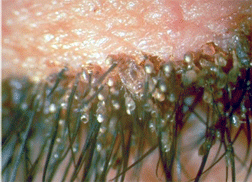
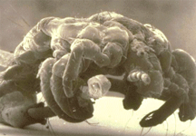
Egg cases, or nits, are attached by female lice to the base of the eyelashes (right). Left, an image of Pthirus pubis taken with an electron microscope.
Images courtesy: Ted Montgomery, O.D.
When the patient returned, the adult organisms, as well as the nits, were removed at the slit lamp with forceps. I successfully removed each nit this waywithout having to remove the lash. Each slid cleanly off without causing the patient any discomfort. The nits and organisms were wiped onto alcohol pads and treated with additional alcohol prior to disposal.
The patient was advised that follow-up treatments would be required because the nits hatch in cycles of seven to ten days.1
At one week, the lashes were examined via slit lamp. No adult organisms or nits were present on the lids O.U., and the crusty appearance of the lid margins had resolved. The patient reported no irritation of the lids.
We scheduled one last follow-up appointment two weeks later. Slit lamp examination again revealed no adult organisms or nits present. The lid margins were normal. No ocular symptoms were reported. The patient confirmed that she had treated other regions of the body. The pupils were dilated using one drop of tropicamide 1% O.U. The cup-to-disc ratio was 0.3/0.3 O.U. The patients maculae, vessels and peripheral retinas were normal.
Discussion

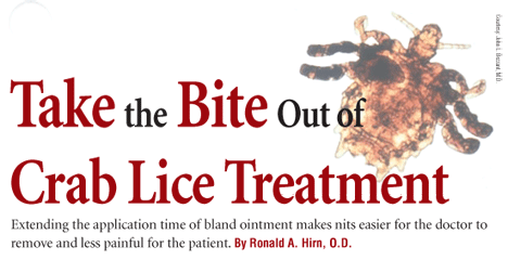
Image courtesy: John L. Bezzant, M.D.
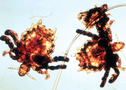
Pthirus pubis, more commonly known as the crab louse, is an external parasite that feeds by sucking blood from the host. These lice prefer to attach themselves (using their middle and hind legs) among the more widely spaced hairs of the pubic region, armpits, and, less commonly, the eyebrows and lashes.
Unlike head or body lice, they are not thought to be vectors for disease, but they can be irritating to the patient, and they tend to remain on their host unless eradicated.2
Lice infesting the human population can be divided into three types: Pediculus humanus corporis (the body louse), Pediculus humanus capitis (the head louse) and Pthirus pubis (the crab or pubic louse). The appearance of the first two are nearly identical, but the last can be distinguished by its broad abdomen and shorter, thicker clawsthese give it the crab-like appearance from which it takes its name.
To feed, the crab louse injects poisonous saliva into the skin. This produces localized scabbing, itching and irritation. A painless bluish discoloration of the skin that does not disappear with pressure may occur around the area of the bite.3 But, sensitivity to the saliva varies, so the patient may present asymptomatic or with symptoms of mild itching.
The process of diagnosis is straightforward: observation of either adult organisms feeding along the lids or egg cases (nits), which the female secures to the lashes with an adhesive. The composition of this proteinaceous adhesive, produced by the female, is similar to that of keratin. It surrounds the egg case, and an opening at the distal end of the case is the only part of the egg not completely encased in this protective sheath.4 Any chemical agents strong enough to denature it would likely also damage the hair follicles. The female lays two to three eggs in a twenty-four hour period, so the lashes may become covered in a very short time.2
Infestation by crab lice is not a new problemPthirus pubis eggs have been found on mummified corpses over 2,000 years old.5 Nor is it an uncommon problem, especially in certain high-risk populations where infestation rates are as high as 52%.6 Be ready to treat and appropriately counsel the patient.
Cases of misdiagnosis as conjunctivitis exist in literature, but this can be avoided by slit lamp examination.7
A variety of methods and pharmacological agents have been reported to successfully remove Pthirus pubis from the eyelashes: yellow mercuric oxide 1%; ammoniated mercuric oxide 3%; cholinesterase inhibitors; fluorescein 20%; and 2.5% pyrethrum cream.1,8-10 But, discussion with a pharmacist may reveal that some of these are rarely available in practice, so they may not be practical.
In this case, I opted for a bland ophthalmic ointment because it is readily available in-office, it is well tolerated by patients, and it has few side effects. Using other ophthalmic ointments, such as mild antibiotic ointments, is acceptablein such cases, the drug vehicle is the more important agent. Antibiotic-steroid combinations can (and perhaps should) be considered if the lid margins are noticeably inflamed, but removal of the offending parasite will typically resolve inflammation.
The use of non-ophthalmic ointments, such as Vaseline, should be avoided because these are likely to result in conjunctival irritation. In many cases, the lashes themselves have to be removed either by epilation or by cutting the lashes at their base, because the nits are adhered so strongly to the lash that the only way to achieve nit removal is to remove the lash as well.11,12 In this case, however, coating the nits with bland ophthalmic ointment over a 24-hour period made them easier to remove. Not enough of these cases are seen in our clinic to provide formal study-based evidence. It would be interesting to see if any other clinicians have repeatable success with this regimen.
Perhaps shorter application times would be as effective, but to reduce the patients waiting time in the office, she was allowed to go home and return. Some practitioners might argue that patients would not want a thick application of ointment to the lids for such a duration, or that they might want the organisms removed immediately. But, I believe that if faced with the painful removal or cutting of the lashes, most patients would at least appreciate an alternative treatment option.
Dr. Hirn attended The Ohio State University. He is in private practice in
1. Sowka J, Gurwood AS, Kabat AG. Handbook of Ocular Disease Management. Jobson Medical Information Inc, March 2000.
2. Weems HV, Fasulo TR. Crab louse, Pthirus pubis (Linnaeus). Available at: http://creatures.ifas.ufl.edu/urban/crab_louse.htm (Accessed June 2007).
3. Buxton PA. The Louse: An Account of the Lice Which Infest Man, Their Medical Importance, and Control.
4. Burkhart CN, Gunning W, Burkhart CG. Scanning electron microscopic examination of the egg of the pubic louse (Anoplura: Pthirus Pubis). Int J Derm 2000 Mar;39(3):201-2.
5. Rick FM, Rocha GC, Dittmar K, et al. Crab louse infestation in pre-Columbian America. J Parasitol 2002 Dec;88(6):1266-7.
6. Imandeh NG. Prevalence of Pthirus pubis (Anoplura: Pediculae) among sex workers in urban
7. Kirschner, M. Pthirus pubis infestation of the eyelashes. JAMA 1982 Jul 23;248(4):428.
8. Kincaid MC. Pthirus pubis infestation of the lashes. JAMA 1983 Feb 4;249(5):590.
9. Mathew M, DSouza P, Mehta DK. A new treatment of phthiriasis palpebrarum. Ann Ophthalmol 1982 May;14(5):439-41.
10. Klaus S, Shvil Y,
11. Yoon KC, Park HY, Seo MS, et al. Mechanical treatment of phthiriasis palpebrarum Korean J Ophthalmol 2003 Jun;17(1):71-3.
12. Ikeda N, Nomoto H, Hayasaka S, et al. Phthirus pubis infestation of the eyelashes and scalp hairs in a girl. Pediatr Dermatol 2003 Jul-Aug;20(4):356-7.

