Unlocking the Genes Behind FECD
A new study may lead to improved screening tools for Fuchs’ endothelial corneal dystrophy.
By Rebecca Hepp, Managing Editor
Researchers from 16 collaborating study sites compared data from more than 5,417 patients, 2,075 of whom were diagnosed with Fuchs’ endothelial corneal dystrophy (FECD), and found new associations between three regions of genetic code, or loci, and the disorder: KANK4, LAMC1 and ATP1B1. They also confirmed the significance of TCF4, which earlier studies have linked to FECD.
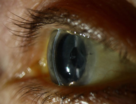 |
| Posterior corneal changes in a patient with FECD and epithelial basement membrane dystrophy. Photo: Christine W. Sindt, OD. |
To study the genes, investigators used corneal tissue from deceased patients, as well as the part of the cornea removed during the transplant procedure in living patients. Immunohistochemistry provided a look at protein expression in the tissue of the living patients.
Taken together, the four loci can predict the risk of FECD with an accuracy of roughly 78%, according to the study. Given no screening tools current exist for FECD, the findings may go a long way to helping clinicians one day better identify patients at risk.
One of the most significant findings, according to the investigators, is that variants of LAMC1 present a significantly higher risk of FECD among women, while TCF4 variants mean a greater risk in men.
These findings hold promise for both better diagnostics and therapeutics for FECD in the future, and ODs should keep an eye on where this research leads.
“While recent advances in transplantation have drastically improved surgical outcomes for Fuchs’ dystrophy, by identifying a genetic basis for the condition, we are able to look toward preventative, rather than curative, management,” says Stephanie Fromstein, OD, assistant professor at the Illinois College of Optometry. “We can move toward in-office testing for those at risk of the condition, as with other point-of-care tests for macular degeneration and Sjögren’s. In-office genetic testing would allow us to identify those at risk much sooner and be more proactive—and successful—in managing their symptoms.”
| Afshari NA, Igo RP, Morris NJ, et al. Genome-wide association study identifies three novel loci in Fuchs endothelial corneal dystrophy. Nature Communications. 2017;8:14898. |
Evidence-Based Pediatric Practice Guidelines
Because eye and vision problems in children have become significant public health concerns, the American Optometric Association (AOA) released a new evidence-based guideline for pediatric eye care.
“An estimated one in five preschool children have vision problems and one in four school-age children wear corrective lenses in this country,” says Andrea Thau, OD, president of the AOA. “Eye health and vision problems in children are significant public health concerns, which is why the new Evidence-Based Clinical Practice Guideline: Comprehensive Pediatric Eye and Vision Examination is so important for doctors of optometry and other health care professionals who treat children.”
The new guideline is designed to increase awareness of the importance of checking children’s eye health, at all ages.
“This provides the only set of evidence-based guidelines for children’s vision care that completely follow the principles outlined in the National Academies of Sciences, Engineering and Medicine’s report Clinical Practice Guidelines We Can Trust,” according to Dr. Thau.
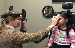 |
| A new guideline can help clinicians succeed with pediatric care. Photo: Kathleen F. Elliot, OD. |
In addition, the guideline provides information to help clinicians:
- Recommend the best interval for in-person comprehensive eye and vision exams for patients from birth to age 18.
- Effectively conduct a full pediatric eye examination, including age-appropriate diagnostic testing procedures.
- Reduce the risk of adverse ocular effects in children.
- Educate patients and their caregivers about eye health and the importance of frequent exams.
The guideline includes new and expanded sections on trauma, myopia, ocular manifestations of child abuse/neglect, color vision deficiency and ultraviolet radiation and blue light protection. The new material highlights how far optometric practice has come since 2002, the AOA said in a press release.1
“What becomes critically important in children is the impact eye care and vision health can have on how well they function in their lives,” says Diane Adamczyk, OD, chair of the AOA EBO Committee, which developed the guideline. “If this guideline heightens the awareness of getting children’s eyes checked, we’ve accomplished our purpose.”| American Optometric Association. AOA releases new evidence-based guideline for pediatric eye care. April 12, 2017. www.aoa.org/news/clinical-eye-care/aoa-releases-new-evidence-based-guideline-for-pediatric-eye-care?sso=y. Accessed April 21, 2017. |
New Therapeutic Target for PDR Identified
A recent study from Massachusetts Eye and Ear found that a small-molecule drug initially developed for cancer therapy led to a significant reduction of abnormal blood vessels in tissue from patients with proliferative diabetic retinopathy (PDR).
“This is a very interesting finding demonstrating the important role of a relatively novel transcription factor—a protein facilitating the conversion of DNA into RNA—implicated in the development of neovascularization seen in several ischemic retinal-vascular diseases, including PDR, neovascular age-related macular degeneration, retinal vein occlusion and retinopathy of prematurity,” explains A. Paul Chous, MA, OD, whose Tacoma, Wash., practice emphasizes diabetic eye care and education.
The first step to the new research was discovering the presence of the transcription factor RUNX1 in abnormal blood vessels, but not normal blood vessels, taken from patients diagnosed with PDR. “RUNX proteins are widely found in multiple species, and their production is exquisitely sensitive to hypoxia as seen in fetal tissue development and tumor growth,” Dr. Chous adds.
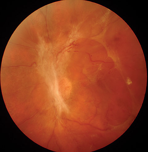 |
| A new therapy for proliferative diabetic retinopathy might be on the way, following new research findings. Photo: A. Paul Chous, MA, OD. |
From there, the investigators treated diseased tissue with the small-molecule RUNX1 inhibitor, leading to a 50% reduction of retinopathy in the preclinical models.
“The fact that a small molecule can hinder the RUNX1 protein is significant because larger molecules, such as those in anti-VEGF agents, face physiologic barriers to absorption that limit their efficacy,” says Dr. Chous. “This opens the possibility to not only topical or oral administration, rather than intravitreal injection, but improved treatment of disease with more targeted therapy and, perhaps, a lower probability of adverse systemic events.”
Current therapies for PDR include laser treatments and anti-VEGF injections, both of which can be inconvenient for patients and come with the possibility, in rare instances, of adverse effects such as retinal hemorrhages, detachments or retinal atrophy, according to the study authors.
“This is the first study to show that blocking the RUNX1 protein is effective at reducing retinal angiogenesis, and it remains to be seen if this strategy is effective, safe and complementary to current anti-VEGF therapies in retinal disease,” says Dr. Chous.
| 1. Lam JD, Oh DJ, Wong LL, et al. Identification of RUNX1 as a mediator of aberrant retinal angiogenesis. Diabetes. April 11, 2017;db161035. [Epub ahead of print]. |
Vitamin A Shows Promise in Retinal Disease
By adding a carbon ring to vitamin A, researchers can induce photoreceptor proteins such as rhodopsin to retain and recycle the designer chromophore molecule, without the help of the retinal pigment epithelial (RPE) cells, according to a new study published in the Proceedings of the National Academy of Sciences.1 Researchers at Case Western Reserve University School of Medicine used the modified form of vitamin A, called locked retinal, to turn on the recycling mechanism and, additionally, activate an important G protein signaling molecule, called transducin, for a longer period of time than naturally occurs.
The revelation puts the biochemistry of vision into the spotlight. “The genesis of vision requires conversion of photons to electrical signals starting in the photoreceptors. In human, this process, known as the “visual cycle,” involves alteration of chromophore 11-cis-retinal to the photoisomerized form known as all-trans-retinal,” says Mohammad Rafieetary, OD, of Charles Retina Institute in Germantown, Tenn.
The reconversion or recycling of these visual pigments occurs in the RPE and depends on healthy RPE and a number of specific proteins. “This fascinating scientific discovery suggests that recycling of chromophores can take place in the photoreceptors themselves, bypassing the RPE and making sensory transduction less dependent on the health of the RPE, which is affected by a number of degenerative diseases,” says Dr. Rafieetary.
RPE plays other significant roles, such as transmitting oxygen and nutrients to the photoreceptors and secreting anti-angiogenic factors to keep a balanced environment for the viability of rods and cones. Therefore, RPE will still remain an essential actor in the pathway. Nevertheless, the discovery may be a significant one for the future of retinal disease treatments.
The research team pioneered the use of another artificial chromophore, 9-cis-retinal, to treat Leber’s congenital amaurosis (LCA), which currently is in Phase II/III of clinical trials, according to Sahil Gulati, a PhD candidate and first author of the current study. “One of the problems of using 9-cis-retinal for the treatment of LCA is the accumulation of spent chromophore that can cause retinal degeneration,” Dr. Gulati says. “The detailed study of the biophysics and biochemisty of locked retinals in this most recent study offers more options to develop treatments for LCA.”
“This study is a revelation that eye care providers need to keep a close eye on,” says Dr. Raffieetary. “One facet we always have to be skeptical of is the development of any scientific discovery to a practical, clinically applied modality, one which has benefits that outweigh the risks.”
While clinicians must be wary of new scientific discoveries, these findings may one day lead to a better way to intervene for debilitating disorders of the retina.
| 1. Gulati S, Jastrzebska B, Banerjee S, et al. Photocyclic behavior of rhodopsin induced by an atypical isomerization mechanism. PNAS. 2017;114(13):E2608. [Epub]. |
In the NewsThe FDA recently approved Genentech’s Lucentis (ranibizumab injection) for the monthly treatment of all forms of diabetic retinopathy (DR). Initially approved to treat DR specificlly in patients with diabetic macular edema (DME), The recent approval came after a Priority Review based on an analysis of the DRCR.net’s Protocol S study. The study compared Lucentis treatment with panretinal laser photocoagulation in DR patients with and without DME, and found the Lucentis group experienced improvements in retinopathy severity.Genentech. FDA approves Genentech’s Lucentis (ranibizumab Injection) for diabetic retinopathy, the leading cause of blindness among working age adults in the United States. April 17, 2017. A new study found the microbial keratitis infection rate for contact lens wearers was higher than for those who had LASIK. While contact lenses were traditionally thought to be safer than surgery, researchers found that soft contact lens wearers, after one year with daily disposables, had fewer cases of microbial keratitis than patients post-LASIK. Masters J, Kocak M, Waite A. Risk for microbial keratitis: comparative metaanalysis of contact lens wearers and post-laser in situ keratomileusis patients. J Cataract Refract Surg. 2017;43(1):67. A new National Institutes of Health (NIH) analysis suggests clinicians should schedule eye exams for patients with Type 1 diabetes based on disease severity, not a typical annual schedule. Adjusting the frequency based on the risk of complications would “result in fewer eye exams at lower cost and quicker diagnosis and treatment of advanced retinopathy,” according to the NIH. National Institutes of Health. Fewer exams and better eye health? Aye-aye, finds Type 1 diabetes study. April 19, 2017. |
