Optometry’s dry eye armamentarium has increased significantly in recent years to include condition-specific artificial tears, anti-inflammatories, better lid disease treatments, amniotic membranes and autologous serum. These have all come about in an effort to treat the growing dry eye epidemic.
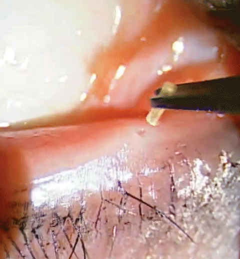 |
| Collagen temporary plugs are inserted into the puncta using forceps. Technically, this is an intracanicular plug. Click image to enlarge. |
With all of these new treatment options available, it’s easy to forget that lacrimal occlusion is a tried-and-true—and often effective—treatment for dry eye. In fact, punctal plugs have been on the market since the 1970s and have become one of the most popular minor procedures in optometry today. One of the many reasons for this is that approximately 25% of tears are lost to evaporation, while the remaining tears drain from the eyes through the minute orifices on the upper and lower eyelids known as the lacrimal puncta.
Maintaining a higher level of moisture on the eye is often achieved through temporary remedies such as artificial tears. But a more permanent method of symptom management can be achieved with the insertion of punctal plugs. The punctal ducts can be occluded with these plugs to help reduce tear drainage and thus retain moisture on the eye, bringing lasting relief to the dry eye sufferer. The growing popularity of this treatment has given way to a large variance of plug sizes, shapes and compositions.
This article examines the process of lacrimal occlusion with both intracanalicular and punctal plugs.
Lacrimal Occlusion
The procedure is a non-pharmacological therapy used to increase the retention of the patient’s own tears. It is primarily used for dry eye syndrome when relief of symptoms is not achieved with first-line treatments, such as ocular lubricants. The inhibition of tear drainage results in increased tear volume and increased contact time of natural tears on the ocular surface. The latter is why lacrimal occlusion may also be used to retain topical medications on the ocular surface.
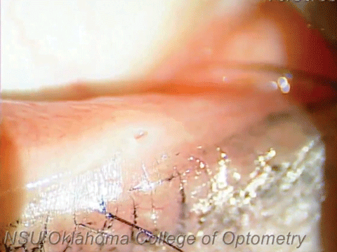 |
| Here you can see the patient’s punctum prior to occlusion. Click image to enlarge. |
Lacrimal occlusion may be temporary, semi-permanent or permanent and can be achieved with the use of intracanalicular plugs, punctal plugs or punctal cautery. Temporary plugs are most commonly intracanalicular and made of collagen. These plugs dissolve and are typically used diagnostically to determine if semi-permanent or permanent lacrimal occlusion should be pursued. Semi-permanent plugs tend to be made of silicone and are considered semi-permanent because they don’t dissolve; however, they can be removed if necessary. Semi-permanent plugs can be punctal or intracanalicular. Permanent lacrimal occlusion is achieved via surgical intervention.
The advantage of intracanalicular plugs is that they may be less irritating to the patient after insertion than punctal plugs; however, it is not easy to confirm their presence after insertion and they can only be removed with lacrimal irrigation. Conversely, punctal plugs remain visible at the surface of the puncta, making it easy to confirm their presence after insertion and making them easy to remove with forceps if needed; however, they can fall out and they may cause minor ocular discomfort.
Patient Selection
Contraindications to lacrimal occlusion include significant inflammation of the ocular surface, inflammation of the eyelids, active infection of the lacrimal system (dacryocystitis), epiphora and silicone allergy (for silicone plugs), or allergy to bovine collagen (for temporary collagen plugs).1,2 It is advisable to ensure there is no abnormal discharge indicative of infection associated with the lacrimal drainage system by applying slight manual pressure to the area.
Punctal occlusion is commonly considered and highly indicated in patients with symptoms of dryness such as ocular irritation/burning sensation, redness and reflex tearing.1,2
Many studies report success improving patient-reported symptoms of dry eye and the procedure is considered both safe and effective when compared to artificial tear use alone.4-6
Young age does not contraindicate the use of punctal plugs. In fact, a study reports that punctal occlusion is safe and effective even for children with symptoms of dry eye—particularly since compliance with other treatment options such as ocular lubricants is difficult in this age group.7
Other indications for punctal occlusion include treatment of assorted ocular surface conditions such as pterygium, pingueculitis, blepharitis, keratitis, corneal ulcers, conjunctivitis, recurrent corneal erosions and other external ocular diseases.1,2 The wound-healing aspect of punctal occlusion is associated with decreased frictional forces against the ocular surface.3
In cases of active ocular inflammation, consider the clinical picture. For example, the increased tear volume attainable through punctal occlusion may not be an acceptable treatment option in many cases of allergic conjunctivitis, as stasis of the offending allergen in the tears may increase a patient’s symptoms.
Procedural Steps
Use of a topical anesthetic agent prior to punctal plug insertion is not necessary, though it can be used if desired. Two suggested methods are to either instill a drop into the conjunctival sac or to hold a cotton-tipped applicator soaked in anesthetic against the punctum for approximately 30 seconds.2
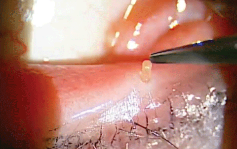 |
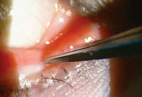
| Once the plug is inserted into the punctum, tap it down using the forceps. It should only take a little pressure for it to fit into place. Click images to enlarge. |
 |
The following steps are common to the insertion of both intracanalicular and punctal plugs (steps are listed assuming the inferior punctum has been chosen to occlude first):
1. Instruct the patient to look up and temporally (away from the inferior punctum).
2. Pull the lower lid down to expose the punctum.
3. Determine the appropriate size plug needed, either by using a punctal gauge or by careful examination, the former being preferred. The punctal gauge can determine the appropriate size (diameter) of plug. If the gauge is too small, there will be no resistance when inserting it; however, if the gauge is too large there will be a significant amount of resistance upon insertion.
4. Dilate the punctum with a punctal dilator if needed.1-2
Following punctal dilation, the following steps are involved in inserting intracanalicular plugs:
1. Using forceps, insert the plug partially into the punctum vertically, then pull laterally, straightening the lacrimal canal, and insert the plug the rest of the way, tilting it towards the nose.
2. Often, the plug can be released from the forceps once it is partially inserted. The tip of the forceps can then be used to push the plug the rest of the way into the punctal opening and into the canaliculus.
3. After insertion, ask the patient to blink a few times to ensure that the plug is in the correct position.
4. Repeat the procedure on the superior punctum if desired. The patient should be instructed to look down and temporally for ease of access to the superior punctum and so the patient is always looking away from where the plug is going to be inserted. To expose the superior punctum, pull the upper lid up.1
If inserting punctal plugs following punctal dilation:
1. Using the applicator that comes with the punctal plug, insert the plug into the punctum until the top of the plug is flush with the lid margin.
2. After insertion, ask the patient to blink a few times to ensure that the plug is in the correct position.
3. Repeat the procedure on superior punctum if desired.
a) The patient should be instructed to look down and temporally.
b) To expose the superior punctum, pull the upper lid up.2
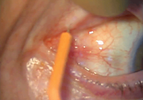 |
| If the plugs fall out, you may consider permanently occluding the puncta. Using an Ellman unit we performed a little punctal cautery to occlude the puncta. This isn’t an option in all states, but it is a procedure that takes only a matter of seconds. Click image to enlarge. |
Plugging In
Both collagen and silicone plugs come in a range of diameters. A common error with plug insertion is failing to use adequately sized plugs or over-dilating the punctum resulting in plug extrusion, or both. A small amount of ocular lubricant can be used on the intracanalicular or punctum plug to aid in insertion through the punctal opening.
Possible complications of lacrimal occlusion include discomfort and irritation at the site of the plug, epiphora, infection, plug migration into the lacrimal drainage system, spontaneous plug extrusion, punctal stenosis, canalicular stenosis, canaliculitis, dacryocystitis and pyogenic granuloma formation.3
The risk of these complications is minimal, but you’ll still want to inform patients of them and advise them to return to the clinic immediately if they experience any symptoms of pain, redness or swelling.
Following plug insertion, follow up in a week or two to reassess the patient’s symptoms and evaluate for side effects. Following punctal occlusion, a patient may continue using ocular lubricants and other medications.
As previously mentioned, lacrimal occlusion may increase the contact time of topical medications on the ocular surface; this may be beneficial in some cases, but you may also consider reducing the dosage of such medications when appropriate.
In the event that collagen plug removal is necessary before the plug dissolves, the intracanalicular plug can be removed by lacrimal saline irrigation—pushing the collagen plug through to the nose or throat.1 A punctal plug can be removed by grasping it with forceps below the exposed plug head and pulling it out of the punctum.
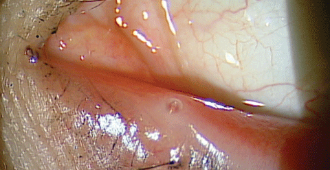 |
| Here you can see the patient’s punctum after occlusion with permanent silicone plugs. |
The most common reasons to remove a plug include local discomfort and epiphora. If the lacrimal occlusion improved overall dry eye without epiphora but was irritating to the patient or spontaneously dislodged, consider punctal occlusion by cautery as an alternative (and permanent) solution. If epiphora is experienced by the patient, a plug that only partially reduces tear drainage may be considered.2
Lacrimal Occlusion in the Literature
Research published in Cornea demonstrates the effectiveness of lacrimal occlusion in a prospective double-masked study, the results of which demonstrated a 94.2% reduction in dry eye symptoms (dryness, watery eyes, itching, burning, sandy/foreign body sensation, fluctuating vision, light sensitivity) and a 93.0% reduction in conjunctival sign/symptoms (redness, discharge) at the eight-week follow-up after progressive occlusion with collagen and silicone plugs.
In contrast, the dry eye and conjunctival symptoms for the control group remained unchanged throughout the eight-week follow-up period. This study also found that eight weeks after progressive lacrimal occlusion, 76.7% of patients were relatively symptom free and 100% of patients were no longer dependent on the daily use of moisturizing agents.8
A 2016 study looked at symptomatic change and fluorescein staining, as well as tear cytokine levels in 29 dry eye patients prospectively. They found significant symptomatic improvement in patient-reported dryness and a decrease in staining on the ocular surface, except inferiorly. They found no decrease in the composition percentage of pro-inflammatory cytokines or MMP-9 by punctal occlusion. Due to the inflammatory nature of ocular surface disease, consider initiating anti-inflammatory therapy at the time of punctal occlusion, as opposed to delaying this treatment.4
In a retrospective literature review, the overall success rate of silicone punctal plug treatment was 76.8% at four weeks and the mean retention time for a silicone punctal plug was 85.1 weeks.9
An observational punctal plug retention and complication study shows an 84.2% three-month retention of silicone plugs, decreasing to 55.8% at two years. It also shows that canalicular stenosis was the most common complication following spontaneous plug extrusion (34.2% at two years); however, patients were asymptomatic to the clinical finding. It is presumed that mechanical stress and accumulation of debris are to blame for the stenosis. Granulomatous proliferation occurred in 3.2% of cases; their review reports that this formation is most likely to occur two to three months after plug insertion. The cause is not completely known, though mechanical injury has been suggested. Two patients in this study experienced plug intrusion as a result of the granuloma, removed under local anesthesia.10
With all this evidence, it is important to keep this simple procedure near the top of your list when treating dry eye patients. Don’t be afraid to reach for the plugs, either canilicular or punctal. Your patients will thank you and tell their friends how you plugged their drain, improved their dry eye, and the dry eye patients will start flowing in.
Dr. Stout is currently completing a family practice residency with an emphasis in ocular disease at Northeastern State University Oklahoma College of Optometry.
Dr. Gillogly is currently completing a cornea and contact lens residency at Northeastern State University Oklahoma College of Optometry.
Dr. Lighthizer is the assistant dean for clinical care services, director of continuing education, and chief of both the specialty care clinic and the electrodiagnostics clinic at NSU Oklahoma College of Optometry.
|
1. Oasis Soft Plug Intranalicular Plug package insert. 2. Oasis Soft Plug Punctum Plug package insert. 3. Bourkiza R, Lee V. A review of the complications of punctal occlusion with punctal and canalicular plugs. Orbit. 2012;31(2):86-93. 4. Tong L, Beuerman R, Simonyi S, et al. Effects of punctal occlusion on clinical signs and symptoms and on tear cytokine levels in patients with dry eye. The Ocular Surface. 2016 Apr;14(2):233-41. 5. Farrell J, Patel S, Grierson DG, et al. A clinical procedure to predict the value of temporary occlusion therapy in keratoconjunctivitis sicca. Ophthalmic Physiol Opt. 2003;23:1-8. 6. Hirai K, Takano Y, Uchio E, et al. Clinical evaluation of the therapeutic effects of atelocollagen absorbable punctal plugs. Clin Ophthalmol. 2012;6:133-8. 7. Mataftsi A, Subbu R, Jones S, Nischal K. The use of punctal plugs in children. Br J Ophthalmol. 2012;96:90-2. 8. Nava-Castaneda A, Tovilla-Canales J, Rodriquez L, et al. Effects of lacrimal occlusion with collagen and silicone plugs on patients with conjunctivitis associated with dry eye. Cornea. 2003;22(1):10-4. 9. Tai M, Cosar C, Cohen E, et al. The clinical efficacy of silicone punctal plug therapy. Cornea. 2002;21(2):135-9. 10. Horwath-Winter J, Thaci A, Gruber A, et al. Long-term retention rates and complications of silicone punctal plugs in dry eye. Am J Ophthalmol. 2007;144(3):441-4. |

