 A 63-year-old Hispanic female presented for an annual eye exam with a chief complaint of blurred vision in her right eye. She has been a patient in the primary eye care clinic for the past 10 years and has always enjoyed excellent visual acuity. During the past two years, however, she began noticing a slow, painless, progressive decrease in her acuity O.D.
On examination, her best-corrected visual acuity measured 20/50 O.D. and 20/20 O.S. One year earlier, her acuity measured 20/30 O.D. and 20/20 O.S. Her ocular history was otherwise unremarkable. Her medical history is significant for systemic hypertension, for which she takes hydrochlorothiazide.
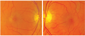
1, 2. Fundus images of our 63-year-old female patient demonstrate retinal whitening around both maculae (O.D. left, O.S. right).
Confrontation visual fields were full to careful finger counting O.U. Ocular motility testing was normal, and her pupils were equally round and reactive to light and accommodation, with no afferent pupillary defect. Her anterior segment exam was unremarkable.
On dilated fundus exam, her optic nerves appeared healthy, with a small cup and good rim coloration and perfusion O.U. We noted subtle fundus changes in both maculae (figures 1 and 2). We also performed spectral-domain optical coherence tomography (SD-OCT, figures 3 and 4) and ordered fluorescein angiography (FA).
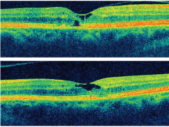
3, 4. A Cirrus HD-OCT (Carl Zeiss Meditech) of the right and left eyes (O.D. top, O.S. bottom). Note the changes found within each macula.
1. What does the OCT show in the patients right eye?
a. Neurosensory retinal detachment.
b. Cystoid space with a retinal drape.
c. Retinal angiomatous proliferation (RAP).
d. Epiretinal membrane with a pseudohole.
2. What does the OCT show in the patients left eye?
a. Full-thickness macular hole.
b. Lamellar macular hole.
c. Epiretinal membrane (ERM), pseudohole.
d. Cystoid macular edema (CME).
3. What is the correct diagnosis?
a. Bulls eye maculopathy.
b. CME.
c. Type 1 macular telangiectasia.
d. Type 2 macular telangiectasia.
4. How should you manage this patient?
a. Observation.
b. Laser photocoagulation O.D.
c. Photodynamic therapy O.D.
d. Intravitreal Avastin (bevacizumab, Genentech) O.D.
For answers, see below.
Discussion
Our patient has macular telangiectasia. This condition may be familiar to readers who regularly read this column, but you may not recognize the condition by this name. Traditionally, we referred to it as juxtafoveal retinal telangiectasia (JRT) or parafoveal retinal telangiectasia. So why the name change? Before answering that question, we need some background information.
J. Donald M. Gass, M.D., and Ray T. Oyakawa, M.D., first characterized idiopathic juxtafoveal retinal telangiectasis in 1982; Dr. Gass and Barbara A. Blodi, M.D., updated this characterization in 1992.1,2 They proposed the use of a classification system that consisted of three groups, each with a subclassification system:
Type 1 JRT. Individuals in this category probably have a mild form of Coats disease. These patients are typically male and have a unilateral presentation that is more often localized to the macula and paramacular area than is seen in Coats disease.1,2 Type 1 JRT patients also tend to be in their mid-30s.
Type 2 JRT. Type 2 patients can be either male or female and usually have a bilateral presentation. This form of telangiectasis is associated with minimal to no lipid exudation and demonstrates superficial retinal crystals in approximately 50% of patients.1,2 Additionally, pigment migration and plaque formation can be seen in the later stages.
Type 3 JRT. This type is exceedingly rare. Capillary occlusions are a hallmark sign.2
Our traditional understanding of JRT has been based on both clinical and FA characteristics. However, advances in recent technology, such as high-speed angiography and SD-OCT, have allowed investigators to study the characteristics of this condition in more detail. So, we now have a much better understanding of the vascular system as well as the structural changes associated with JRT.
This, in turn, led Lawrence A. Yannuzzi, M.D., and associates to devise a simplified JRT classification system. In this new classification system, they adopted the name idiopathic macular telangiectasia and identified two distinct groups.
Group 1 was categorized as macular aneurysmal telangiectasia (MAT), and group 2 was categorized as macular perifoveal telangiectasia (MPT).3,4
Newer imaging technologies have helped identify some interesting differences between the two groups. Patients in group 1 are still considered to fall within the spectrum of Coats disease. These patients are more likely to have profound vascular changes, with more obvious aneurysmal dilations and prominent cystic changes within the macula.
These extensive macular cystic changes are not seen in group 2 patients. Instead, patients in group 2 have a central lamellar cyst, which the retina drapes over, that is visible on OCT. This finding has become the hallmark diagnostic sign for a group 2 patient. Group 2 patients also demonstrate a loss of retinal transparency; smaller, subtler telangiectatic changes within the capillaries; and retinal pigment epithelial (RPE) changes not seen in group 1 patients. The changes within the RPE explain why group 2 patients can develop choroidal neovascularization in the conditions later stages.3,4
Our patient has type 2 macular telangiectasia. On clinical exam, the retinal whitening seen around the maculae O.U. signals a loss of retinal transparency. There are fine telangiectatic retinal vessels, which are essentially invisible on clinical examination. The OCT shows the lamellar cysts in each eye, with draping of the inner retinal layers over them.
Interestingly, OCT of our patient reveals a break in the roof of the inner retina O.S., which has created a lamellar hole. Surprisingly, the patient still sees 20/20 O.S. despite these macular changes. We considered an Avastin injection for her right eye, but the patient decided that her symptoms were not bad enough to justify treatment.
Even as we begin to better understand macular telangiectasia, there are still questions that remain unanswered. More effective treatments are still being sought out, and investigators are still not sure what causes it. Some have proposed that systemic factors may play a role, while others suspect a genetic link.
A multi-center clinical trial has been established to determine the natural history of macular telangiectasia and determine if the condition has a genetic link.5 The researchers also hope to explore more effective treatments. The study, called the MacTel Project, is the only international project devoted entirely to the study of macular telangiectasia. More information about this study can be found at www.mactelresearch.org.
Retina Quiz Answers: 1) b, 2) b, 3) d, 4) a.
1. Gass JD, Oyakawa RT. Idiopathic juxtafoveolar retinal telangiectasis. Arch Ophthalmol 1982 May;100(5):769-80.
2. Gass JD, Blodi BA. Idiopathic juxtafoveolar retinal telangiectasis: update classification and follow up study. Ophthalmology 1993 Oct;100(10):1536-46.
3. Yannuzzi LA, Bardal AM, Freund KB, et al. Idiopathic macular telangiectasia. Arch Ophthalmol 2006 Apr;124(4):450-60.
4. Chew E, Gillies M, Bird A. Macular telangiectasia: a simplified classification. Arch Ophthalmol 2006 Apr;124(4):573-4.
5. The MacTel Project. What is MacTel? Available at: https://web.emmes.com/study/mactel/donor/donor_brochure.pdf (Accesed January 11, 2009).
