 History
A 66-year-old white male presented with a chief complaint of poor vision in his right eye that had persisted for one week. The patient explained that he knew he had a “lazy right eye” that required surgical correction when he was younger. Although the patient admitted he never saw well with his right eye, he insisted that his vision recently decreased significantly.
When the left eye was covered, he described the vision in his right eye as if he were looking through Vaseline-smeared glass (which was particularly noticeable in the left field). Further, the patient remarked that a previous eye care provider informed him that his right retina had an “unusual appearance.”
He denied using any prescription medications and reported no known allergies.
Diagnostic Data
His best-corrected entering visual acuity was 20/400 OD and 20/30 OS at distance and near (refraction and pinhole testing yielded no improvement). His pupils were equal, round and reactive to light, with a grade II afferent defect OD. Extraocular muscle movements were full OU.
Confrontation fields revealed a left hemifield loss in the right eye. Slit lamp examination uncovered normal anterior segment structures and anterior chambers. His IOP measured 18mm Hg OU.
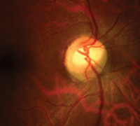
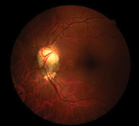
Fundus photographs of our 66-year-old patient who presented with poor vision in his right eye (OD left, OS right). What do you notice?
Discussion
Additional screening procedures included automated visual fields, brightness and color testing (with specific attention to ruling out red desaturation). We photodocumented the posterior poles and obtained a frequency-doubling field.
The diagnosis in this issue is bilateral morning glory syndrome (MGS) with evidence of retinal detachment bordering the right macula.
Congenital abnormalities of the optic disc can be unilateral or bilateral, and can spare visual function or impair it mildly to severely.1-3 The “excavated optic disc abnormalities” include MGS, optic disc coloboma and staphyloma.1
Optic nerve hypoplasia is distinguished by a small papilla that is frequently accompanied by a peripapillary ring (the double ring sign).1 Patients with the morning glory anomaly typically exhibit a tilted nerve with areas of atrophy throughout the disc.1
MGS has been associated with craniofacial anomalies, basal encephalocele and retinal detachment secondary to slit-like breaks at the margin of excavation.2,3 MGS and basal encephalocele always should be investigated when midline deficiencies are accompanied by the ophthalmic signs of strabismus or poor vision.2 The pathogenesis of MGS and basal encephalocele are unknown; however, they are thought to be linked to issues that transpire during the fifth week of embryonic development.
Computed tomography and magnetic resonance imaging should be performed to rule out the presence of any additional lesions, and a complete hormone screening should be ordered to exclude pituitary deficiency.2
Optical coherence tomography may be beneficial in the detection of these subtle, slit-like breaks at the margin of excavation in MGS patients. Further, the technology can provide guidance toward the requirement of subsequent surgeries and may prove helpful in confirming postoperative closure status.3
We immediately referred the patient to a retinal specialist for vitrectomy surgery. His postoperative acuity improved to 20/80 OD. We anticipated cataractogenesis following the vitrectomy, and educated the patient that he likely will require cataract surgery within one year.
1. Dutton GN. Congenital disorders of the optic nerve: excavations and hypoplasia. Eye. 2004 Nov;18(11):1038-48.
2. Chen CS, David D, Hanieh A. Morning glory syndrome and basal encephalocele. Childs Nerv Syst. 2004 Feb;20(2):87-90.
3. Ho TC, Tsai PC, Chen MS, et al. Optical coherence tomography in the detection of retinal break and management of retinal detachment in morning glory syndrome. Acta Ophthalmol Scand. 2006 Apr;84(2):225-7.
