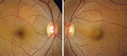
 A 28-year-old Hispanic female presented with blurred vision in her left eye that had persisted for three weeks. Her local optometrist noted some suspicious findings in her retinae, and recommended that she visit Bascom Palmer for a more extensive evaluation. Her ocular history was unremarkable. She was in good health, and she was 33 weeks pregnant. She regularly took prenatal vitamins.
On examination, her best-corrected visual acuity measured 20/20 O.D. and 20/30 O.S. Extraocular motility testing was normal. Her confrontation visual fields were full to careful finger counting O.U.
Her pupils were equally round and reactive, with no afferent pupillary defect. Amsler grid testing was normal in each eye. Further, the anterior segment examination was unremarkable.

1, 2. Our patient’s eyes (O.D. left, O.S. right) demonstrate unusual changes scattered throughout the posterior poles and the arcades.
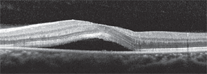
Her vitreous was clear on dilated fundus exam. She had moderate-sized cups with good rim coloration and perfusion in both eyes. The Fundus examination of the right macula was remarkable for some obvious retinal changes seen superior to the macula (figure 1). The fundus examination of her left eye also showed more subtle retinal changes involving the macula (figure 2). Additionally, we performed spectral domain optical coherence tomography (SD-OCT) imaging (figure 3).
3. A horizontal SD-OCT slice through our patient’s left macula. What do you see?

Take the Retina Quiz
1. What do the retinal changes seen in both eyes represent?
a. Multiple serous retinal detachments.
b. Multiple retinal pigment epithelial detachments (PEDs).
c. Localized choroidal neovascularization (CNV).
d. Polypoidal choroidal lesions.
2. What additional testing is necessary for this patient?
a. Fluorescein angiogram.
b. Visual fields.
c. Blood pressure testing.
d. Pattern electroretinogram (ERG).
3. What is the likely diagnosis?
a. Idiopathic retinal pigment epithelial detachments.
b. Idiopathic central serous choroidopathy (ICSC) associated with pregnancy.
c. Toxemia of pregnancy.
d. Polypoidal choroidal vasculopathy.
4. How should this patient be managed?
a. Anti-vascular endothelial growth factor therapy.
b. Photodynamic therapy (PDT).
c. Laser photocoagulation.
d. Observation; return in two to three weeks.
For answers, see below.
Discussion
Our patient had bilateral central serous retinal detachments; the detachment in her left eye involved the macula. Fortunately, her right macula was unaffected because the serous detachment was located superior nasal to the macula. But, because the detachment in her right eye was more sharply demarcated and “blister-like” than that in her left, it could possibly represent a pigment epithelial detachment. An OCT scan through the detachment would confirm its location. Unfortunately, however, the scan was performed through the macula, not the lesion.
ICSC typically affects healthy males between 20 and 45 years of age. In fact, men are five to 10 times more likely to develop ICSC than women.1 Stress seems to play an important factor in the development of ICSC. Also, the condition commonly occurs in individuals with “type A” achievement-oriented personalities.1
So, how can we confirm that our patient has ICSC? Clearly, she is not male. And, she did not report any unusual stress. But, interestingly, a subset of ICSC can occur in pregnant females.
Initial reports of central serous retinopathy in pregnancy surfaced in 1974.2,3 But, by 1993, just 14 cases had been reported.1 ICSC may occur during any trimester, and most patients demonstrate complete resolution of the detachments near the end of pregnancy or within a few months after delivery. Previously, the literature suggested that ICSC secondary to pregnancy was exclusive to non-whites; however, it is now believed that the condition may present in individuals of any race. Additionally, subretinal fibrinous exudates develop in approximately 90% of ICSC cases associated with pregnancy compared to just 20% of cases not related to pregnancy.4
Our patient clearly demonstrates subretinal fibrinous exudates. The fibrinous exudate is seen as lighter-colored punctate spots within the serous detachments. Some of them even have a drusenoid appearance.
The pathogenesis of ICSC is still not completely understood. It has been associated with increased cortisol levels in patients who take corticosteroids for systemic disease or have received organ transplants. In pregnancy, a woman experiences increased levels of free circulating endogenous cortisol, which likely plays an important role in the development of ICSC.2-4
It is important to monitor blood pressure in pregnant women who present with serous retinal detachments or demonstrate suspicious retinal findings. Pregnancy-induced hypertension, or toxemia, can cause serious systemic and ocular consequences. Mild toxemia (preeclampsia) affects roughly 5% to 10% of pregnant women. Severe toxemia, however, can cause significant health problems, including organ damage. Our patient’s blood pressure measured 112/70mm Hg, which was not high.
The recommended treatment for both conventional and pregnancy-related ICSC is observation, because most patients spontaneously improve within six months without medical intervention.1-4 We instructed our patient to return in three weeks for a follow-up examination. At that visit, her condition was improved; however, she still had a small amount of serous fluid in each eye. We saw the patient one month after her delivery, and the retinal detachments had resolved completely.
Retina Quiz Answers: 1) a; 2) c; 3) b; 4) d.
1. Gass JD. Stereoscopic Atlas of Macular Disease: Diagnosis and Treatment, 4th ed. St. Louis: Mosby, 1997:52-70.
2. Chumbley LC, Frank RN. Central serous retinopathy and pregnancy. Am J Ophthalmol. 1974 Feb;77(2):158-60.
3. Sunness JS, Haller JA, Fine SL. Central serous chorioretinopathy and pregnancy. Arch Ophthalmol. 1993 Mar;111(3):360-4.
4. Gass JD. Central serous chorioretinopathy and white subretinal exudation during pregnancy. Arch Ophthalmol. 1991 May;109(5):677-81.

