 A 53-year-old black male presented complaining of sudden-onset red floaters in his right eye. His previous ocular history was remarkable for non-proliferative diabetic retinopathy as well as mild cataracts in both eyes. His systemic history was remarkable for type 2 diabetes, hypertension, hypercholesterolemia and congestive heart failure. In addition, he was recently diagnosed with obstructive sleep apnea (OSA). The patient reported smoking four cigarettes per day. His hemoglobin A1C was 8%. Systemic medications included Norvasc (amlodipine besylate, Pfizer), digoxin, insulin, aspirin, Accupril (quinipril, Pfizer) and Lipitor (atorvastatin, Pfizer). The patient measured 5 feet 9 inches tall and weighed 215 pounds. He had an appointment to see his internist later that week.
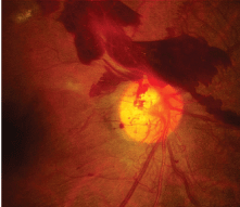
The patients best-corrected distance acuity was 20/30 O.D. and 20/40 O.S. Biomicroscopy revealed bilateral grade 1 cortical cataracts. Intraocular pressure was 15mm Hg O.U. Dilated funduscopy revealed proliferative diabetic retinopathy and clinically significant macular edema in each eye. Vitreous, pre-retinal and retinal heme was present in the right eye. His blood pressure measured 150/90mm Hg.
A summary report was dictated and sent to the patients internist. We referred the patient to a retina specialist for appropriate treatment. His OSA was treated using continuous positive airway pressure (CPAP).
The patients A1C and blood pressure measurements suggest less than optimal control of his diabetes. We know that these factors may have an adverse affect on his retinopathy. Could his sleep apnea be an aggravating factor as well?
Could this patients sleep apnea be causing his diabetic retinopathy to progress more rapidly?
Sleep Disorders Affect Health
There are five stages of sleep that cycle continuously throughout a single nights rest. Stages one through four are known as non-rapid eye movement (non-REM) sleep. The fifth stage is rapid eye movement (REM). A complete sleep cycle lasts about 90 minutes.1
Adequate sleep is essential for proper immune system, nervous system, physical and mental function. However, some 70 sleep disorders affect 40 million people in the United States.1 These disorders are classified into three categories: lack of sleep (insomnia), excessive sleep (narcolepsy) and disturbed sleep (OSA, REM sleep behavior disorder, restless leg syndrome and periodic limb movement disorder).
Obstructive sleep apnea is a common chronic disorder that causes one or more pauses in breathing (apneas) or shallow breaths during sleep.2,3 Loud snoring, choking and gasping during sleep are the most common signs of OSA.3
An estimated 12 million or more American adults have OSA.3 It is often found in patients with obesity, diabetes or cardiovascular disease.2,3 Men are more likely to have OSA than women. In fact, one out of 25 middle-aged men and one out of 50 middle-aged women has OSA.
The consequences of OSA include excessive daytime sleepiness, impaired cognitive functioning, increased cardiovascular morbidity (coronary artery disease/myocardial ischemia), cerebrovascular accident and overall mortality.2
What Causes OSA?
OSA is caused by recurrent obstruction of the airway during sleep. The pharynx (throat) is separated into the nasopharynx (connects the back of the nose to the back of the mouth), the oropharynx (connects the mouth to the top of the throat) and the hypopharynx (sits behind and on either side of the larynxthe voice box).1 Anatomic narrowing in the nasopharynx and oropharynx causes increased resistance to airflow. Sleep-related reduction in muscle tone may also further compromise these areas.2 Tonsillar hypertrophy, obesity (causing wide neck circumference), consuming alcoholic beverages or using sedatives at bedtime may contribute to reducing the diameter of any portion of the upper airway.
In OSA, ventilation is reduced (hypopnea) or absent (apnea) for several seconds until upper airway muscle tone increases. Ventilation is then restored until deeper sleep resumes and muscle tone once again diminishes, causing the cycle to repeat.2 Breathing pauses can last from seconds to minutes and can occur five to 30 times per hour.3 OSA is more severe in the supine position and more apparent during REM sleep.2
How is OSA Diagnosed?
The diagnosis of OSA is based on a patients medical history, physical examination and test results from a sleep study.3 The most common study for diagnosing OSA is overnight polysomnography (PSG), conducted in a sleep laboratory.2,3 PSG is painless; sensors are placed on the scalp, face, chest, limbs and finger.3 This allows for monitoring of snoring, pulse oximetry, electrocardiogram (EKG), muscle tone, eye movements and electroencephalogram (EEG).2
The physiological changes that may occur in OSA include systemic and pulmonary hypertension, dysrrhythmia, reduced cerebral blood flow, increased left ventricular afterload and decreased cardiac output.2
Complications of OSA
OSA often coexists with diabetes. Obesity is a risk factor for both type 2 diabetes and OSA. Patients with OSA typically have a body mass index (BMI) greater than 25 kg/m2 and a neck circumference of more than 17 inches in men and more than 16 inches in women. Studies have also found a link between OSA and hypertension. OSA may aggravate diabetic retinopathy, secondary to nocturnal hypertension and hypoxemia.2,4,5
Ocular complications of OSA include floppy eyelid syndrome, glaucoma (primary open-angle and normal tension), papilledema, and non-arteritic anterior ischemic optic neuropathy (NAION).4,6,7
Managing OSA
Management of OSA is aimed at restoring regular breathing and relieving symptoms, such as loud snoring and daytime sleepiness.3 Strategies include increasing physical activity, sleeping in a non-supine position, and avoiding alcohol, hypnotics and narcotics before bedtime. Other treatments range from oral appliances to moderate intervention, such as CPAP. More radical approaches include surgical removal of anatomic obstructions.
In CPAP therapy, patients are fit with a nasal or nasal/oral mask that allows inflow of pressurized air to prevent collapse of the airway during sleep.
CPAP is effective at treating the majority of cases of sleep apnea for patients that regularly use this therapy. Between 65% to 75% of patients tolerate it well.8
However, some individuals have difficulty tolerating CPAP. A feeling of claustrophobia and a perceived inconvenience may lead to poor compliance.2 Treatment with CPAP may also cause side effects, such as a dry or stuffy nose, irritated skin, dry eyes and headache.3 Other patients report an uncomfortable air pressure sensation in the nose or throat. For the latter patients, several types of masks have been developed that may help.
In this patients case, CPAP therapy improved his OSA symptoms. For his advanced proliferative retinopathy, vitrectomy in the left eye and laser therapy in both eyes were the best options. The patient underwent pars plana vitrectomy (PRP) with endolaser photocoagulation in each eye. The patient also had grid laser photocoagulation therapy in each eye for his macular edema. This treatment, along with the CPAP, improved his ocular health and vision.
Eye care professionals need to be able to recognize symptoms of OSA and refer those patients to a sleep specialist.4 Patients with diabetes who have signs or symptoms of OSA should be evaluated for this condition because OSA may cause the retinopathy to worsen.5 By the same token, sleep medicine specialists should refer their patients with OSA for an appropriate ophthalmic evaluation.
1. Kernich CA. Sleep disorders. Neurologist 2002 Mar;8(2): 129-30.
2. Boyer S, Kapur V. Obstructive sleep apnea: Its relevance in the care of diabetic patients. Clin Diabetes 2002 July 1;20(3): 126-32.
3. National Heart, Lung, and Blood Institute. Diseases and Conditions Index Web site. Sleep apnea. 2009 Mar. Available at: www.nhlbi.nih.gov/health/dci/Diseases/SleepApnea/
SleepApnea_WhatIs.html (accessed April 7, 2009).
4. Abdal H, Pizzimenti JJ, Purvis CC. The eye in sleep apnea syndrome. Sleep Med 2006 Mar;7(2):107-15.
5. Sinclair SH,
6. Mojon DS, Goldblum D, Fleischhauer J, et al. Eyelid, conjunctival and corneal findings in sleep apnea syndrome. Ophthalmology 1999 Jun;106(6):1182-5.
7. Mojon DS, Mathis J, Zulauf M, et al. Optic neuropathy associated with sleep apnea syndrome. Ophthalmology 1998 May;105(5):874-7.
8. Giles TL, Lasserson TJ, Smith BH, et al. Continuous positive airways pressure for obstructive sleep apnoea in adults. Cochrane Database Syst Rev 2006 Jul 19;3:CD001106.
