Glaucoma continues to be a captivating area of study, with groundbreaking research on a new instrument for measuring visual evoked potentials, glaucoma medications and the impact of central corneal thickness (CCT) on disease risk and progression. Here are a few highlights from this years Association for Research in Vision and Ophthalmology (ARVO) meeting.
Central Corneal Thickness
Research presented at this years meeting confirms findings of the Ocular Hypertension Treatment Study (OHTS) and highlights the growing importance of CCT in the management of glaucoma.
For example, researchers at the Indiana University School of Medicine found that differences in CCT between the eyes of a single patient can have a significant impact on progression of glaucoma in each eye.2478 The retrospective study looked at the records of 69 patients who have primary open-angle glaucoma. Patients were excluded if they had vision worse than 20/40, corneal dystrophy or disease, retinal pathology or macular disease. When examining mean deviation of visual field tests and pattern standard deviation, the researchers found a significant correlation between thinner corneas and an increased severity of glaucoma.
CCT also appears to be a powerful clinical factor in assessing structural damage during the initial examination of a normal-tension glaucoma patient, concludes a multi-center study conducted in Korea and Australia.1298 Researchers analyzed 75 eyes of 75 normal-tension glaucoma patients with early to moderate visual field defects and localized retinal nerve fiber layer defects. Thinner corneas were significantly associated with increased horizontal cup-to-disc ratios. CCT should always be considered when assessing normal-tension glaucoma patients, the researchers say.
Medications
A retrospective review of patient charts at a Chicago hospital found that glaucoma patients who responded successfully to treatment in one eye with only Travatan (travoprost, Alcon) or Lumigan (bimatoprost, Allergan), showed a statistically significant decrease in IOP in the contralateral eye.3767 Twenty-two patients underwent monocular trials with travoprost, and 17 underwent monocular trials with bimatoprost. In the treated eyes, travoprost reduced IOP 33.3% on average, and bimatoprost caused a 35.3% drop. In the contralateral eye, travoprost caused a 7.1% reduction in IOP, while bimatoprost produced an 11.3% drop.
Also, a prospective, randomized, study in Rome found that patients can minimize hypertrichosis and periocular skin pigmentationtwo possible side effects of bimatoprostby wiping the eye with a highly-absorbent pad after administering the drop.2457
Research at this years conference looked at instrumentation, including comparisons between:
Dynamic contour tonometry and Goldmann applanation tonometry. Differences in subject age and CCT skew the readings of the Goldmann applanation tonometer to a greater degree than they do the Pascal dynamic contour tonometer, according to a study in London.4838 Researchers looked at 130 eyes in 130 patients, and used Bland-Altman plots-mean difference to determine true IOP. They concluded that between the youngest and oldest eyes, and between the thinnest and thickest corneas, age and CCT accounted for a variation of 3.0mm Hg between the two devices. The effects were independent of each other.
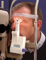 |
| Readings of the Pascal dynamic contour tonometer are less skewed by age and central corneal thickness than Goldmann applanation tonometry, according to one study presented at this years meeting. |
Scanning laser polarimetry (SLP), confocal scanning laser ophthalmoscopy (CSLO) and stereo photographs. In a study conducted in the Netherlands, researchers measured one eye in 40 healthy subjects, 48 glaucoma patients, and six patients who had ocular hypertension with SLP using the GDxVCC (Variable Corneal Compensator) and CSLO using HRT- I (Heidelberg Retina Tomograph).4809 (HRT-II, the newst iteration of the HRT, was not tested in this study.) They also obtained simultaneous stereoscopic optic-disc photographs for analysis by four glaucoma specialists, four general ophthalmologists, four residents in ophthalmology and four optometrists. Automated analysis of measurements with SLP and CLSO had a higher diagnostic accuracy than classification of stereoscopic optic-disc photographs by glaucoma specialists, the researchers found. The intra- and interobserver agreement for optic-disc analysis was only moderate to good.
FDT and SWAP perimetry. Researchers in Italy attempted to evaluate the effectiveness of frequency-doubling technology (FDT) and short wavelength automated perimetry (SWAP).2486 Study subjects included 23 patients with glaucomatous optic nerve head and retinal nerve fiber layer (RNFL) defects but normal standard automated perimetry (SAP). Patients were followed for five years and then retested with the same procedures.
At the start of the study, 70% had an abnormal FDT, and 26% had an abnormal SWAP. At the five-year follow-up, 52% developed glaucomatous visual field defects on SAP. Every one of the 52% (12 patients) had an abnormal FDT and abnormal SWAP at the five year follow-up. Eleven had abnormal FDT at inclusion; five had abnormal SWAP at inclusion. Defects on both FDT and SWAP might preceed the onset of SAP changes in early glaucoma, although FDT might detect progression of glaucoma earlier than SWAP, the researchers concluded.
Meanwhile, a multicenter study in New York tested a new noninvasive visual evoked potential device.3758 The instrument is designed to detect dysfunction in divisions of the magnocellular pathway, which previous studies have shown to be related to glaucoma. A potential advantage: objective measurement of visual function.
Researchers hoped to equal the results achieved by previous, more labor-intensive versions of this technology. So, they collected data from 23 participants12 open-angle glaucoma patients, three glaucoma suspects, two ocular hypertensives and six age-matched controlswho had a minimum visual acuity of 20/30. In 10% contrast stimulation conditions, the device revealed neural deficits in 11 of 14 glaucomatous eyes that had 20/20 visual acuity. Preliminary analysis of the data yielded accuracy estimates ranging from 79% to 96%, consistent with results achieved using earlier techniques. The authors hope the new instrument will allow quick, early detection of central visual deficits in glaucoma patients.
Blood Flow
Glaucoma patients show a significantly smaller increase in optic nerve head blood flow than healthy individuals in response to flicker stimulation, according to a study conducted in Montreal.5731
Researchers continuously monitored nine healthy patients and 11 glaucoma patientsthe glaucoma patients were being treated with topical hypotensive therapyusing laser Doppler flowmetry to detect blood flow changes on the superior-temporal neuroretinal rim. A green light flickering at 16Hz through the fundus camera while monitoring patient fixation.
Blood flow increased 64% in normal subjects but only 7% in glaucoma patients. The authors believe that, despite IOP-lowering treatment, some underlying vascular disturbances related to glaucoma remain unchanged.
Dr. Cole is in private practice in Bridgeton, N.J., and is an assistant professor at Pennsylvania College of Optometry.
1298. Kim D, Choi H, Pye DC. Central corneal thickness as a risk factor for glaucomatous damage in normal-tension glaucoma.
2457. Parravano M, Centofanti M, Manni G, et al. Preventing cosmetical side-effect of prostamide topical therapy.
2478. Rogers DL, WuDunn D, Cantor LB, Catoira YM. Comparison of central corneal thickness and visual field loss within the same patients with open angle glaucoma.
2486. Rossetti L, Fogagnolo P, Mazzolani F, et al. Detection of early glaucoma with FDT and SWAP: the results of a longitudinal study.
3758. Zemon VM, Tsai JC, Chen CM, et al. A novel electrophysiological instrument for rapid and objective assessment of glaucomatous damage.
3767. Golden AM, Shah M, Emerick G. Contralateral IOP effect of travoprost and bimatoprost.
4809. Reus NJ, Lemij HG. Accuracy of GDx VCC, HRT I, and clinical assessment of stereoscopic optic disc photographs for diagnosing glaucoma.
4838. Kotecha A, White ET, Shewry JM, Garway-Heath DF. The relative effects of corneal thickness and age on Goldmann applanation tonometry and dynamic contour tonometry.
5731. Wajszilber M, Descovich D, Deveault A, Lesk M. Changes in optic nerve head blood flow in treated glaucoma patients versus normal subjects during flicker stimulation.
