 |
 |
The majority of surgical advancements tend to be subtle and relatively inconspicuous to both patients and their optometrists. One recent advancement, on the other hand, is as big and obvious as it gets. 3D technology in the surgical suite is really straight out of the movies.
Stereoscopic high-definition surgical visualization systems are not brand new in medicine, but they are now starting to permeate various areas of eye surgery. They have been more widely used in cataract and glaucoma surgeries in the last several years, but as this month’s Surgical Minute video shows, there are pioneering applications in retina surgery as well. This is not a trivial barrier to cross, as retina surgery is arguably the most delicate ocular surgery out there.
The system uses a three dimensional camera attached to the surgical microscope, and a high-resolution plasma television is placed at the end of the bed. The surgeon and support staff can use 3D glasses to watch the surgery in real time with enhanced stereopsis.
Benefits
Support staff who can see the surgeon’s viewpoint can also better anticipate how to help. This video illustrates how a surgeon can successfully perform even the most complex and delicate of retinal surgeries, such as vitrectomy and membrane peel in an eye with proliferative retinopathy. Complicated steps, such as silicone oil tamponade and bimanual manipulation, can been seen in this video.
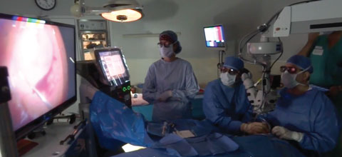 |
| Anyone in the surgical suite wearing 3D glasses will be able to appreciate the stereopsis and the depth of field that the surgeon’s seeing. |
For the surgeon, a 3D heads-up display provides significant improvements to traditional surgery. It allows surgeons better visual and ergonomic comfort, as they do not have to bend over and fixate through a microscope. It can also provide enhanced depth of field, leading to more precise surgeries.
Future Advancements
Developing heads-up technologies will provide even more significant surgical refinement, as researchers are testing the use of real-time OCT and other ocular imaging that can be displayed on the screen simultaneously or overlaid on the real-time image. This will give surgeons potentially endless data displayed in their surgical view.
Three dimensional real-time imaging also represents an innovative new platform for education. Surgeons and support staff in training are able to fully appreciate the surgical procedure through the surgeon’s view. New surgeons in training can also be evaluated and coached by peripheral mentors.
But the educational opportunities don’t end in the OR. Watching three dimensional surgeries affords optometrists a better understanding of techniques and in vivo anatomy. It also reinforces the complexity and delicate nature of ocular surgery. Some surgeons even find this technology beneficial for educating patients about ocular surgery.
3D surgery represents a significant departure from the standardized methods of imaging ocular surgery, so it may come as no surprise that adoption may be slow. Yet, as this technology continues to evolve, we can expect its growth in popularity for surgeons, and better outcomes for patients.