This patient has Charles Bonnet syndrome (CBS), a condition in which patients experience complex visual hallucinations, often pleasant in nature, that cannot be explained by any disturbance of thought or the presence of a psychiatric disorder.1-3
 |
As primary-care clinicians, we can educate ourselves and our patients (particularly those who already have or are at risk for CBS) about the condition. This, in turn, may make patients who have CBS less reluctant to tell us about their visual hallucinations. Here, we will discuss the epidemiology and history of CBS, its pathogenesis, diagnosis and management.
Epidemiology
CBS most often occurs in elderly individuals who are usually mentally healthy.5 Patients are always fully aware that the visual hallucinations they experience are not real.6
The mean age of incidence is about 57 to 80, but CBS also has been reported in children who have rapid vision loss.2,7 The predilection for the elderly merely reflects the increased incidence of sudden, profound vision loss in that age group.2 Although most CBS patients have vision loss, this is not required to diagnose the condition.5
We use the term Charles Bonnet Plus (CBS Plus) to describe clinical cases in which visual hallucinations are associated with other cognitive, psychopathologic or neurological disorders.8 Patients who have CBS Plus have cognitive impairment, while CBS patients are wholly mentally intact. The difference between CBS Plus and mental illness is that patients who have CBS Plus have both cognitive and visual deficits that contribute to their state of sensory deprivation.
CBS is named after Charles Bonnet (1720-1793), a Swiss naturalist, biologist and philosopher who was the first person to use a scientific approach in the study of hallucinations and related phenomena.3
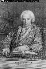 |
| Charles Bonnet was the first person to use a scientific approach in the study of hallucinations and related phenomena. |
Mr. Lullin, a magistrate, remained cognitively intact and fully realized that his visions were fictions of his brain.2,11 He told his grandson that he was not frightened by these images but rather intrigued and amused by them.3,12 Ironically, Dr. Bonnet also experienced these visual hallucinations as his own vision worsened near the end of his life.1,6 (No source can be found that reveals the cause of Bonnets visual deterioration.)
Pathogenesis
Bilateral simultaneous or sequential vision loss from any cause can result in visual hallucinations. (See A Look at Visual Hallucinations) AMD is the most com-mon triggering pathology of CBS, although it also has been associated with pathology at any level of the visual system.12,13 This includes any condition that affects the visual pathway from the eyeball, optic nerve and visual radiations of the brain to the occipital cortex. These visual hallucinations are often referred to as release hallucinations because they arise from or are released in a visual cortex that no longer receives the incoming visual sensory impulses that usually filter out nonvisual stimuli.14
This release phenomenon is part of the sensory deprivation theory that explains CBS.12,15 The vision loss produces a state of relative sensory deprivation that allows these images to be released into conscious perception.16 This theory has also been called the phantom-vision theory and has been compared with phantom-limb syndrome. Phantom-limb syndrome occurs in a patient who experiences sensory sensations, such as burning at the soles of her foot, even though her leg was amputated.2,16,17
Cortical input from other areas of the brain, such as memory association areas, also may fill in for the lack of sensory input from the eyes. Indeed, CBS patients who undergo vision-restoring surgery (cataract surgery, macular translocation, etc.) no longer experience visual hallucinations.12,18-20
The degree of visual impairment is important in the formation of these release hallucinations. CBS occurs more frequently in patients who have a higher degree of visual impairment and bilateral pathology.2,19,20 Patients do not experience unilateral hallucinations unless they perceive the images more in one eye than in the other. Release visual hallucinations usually occur in patients who have a visual acuity of 20/60 or worse in the better eye.2,19,20
Most visually impaired individuals do not experience visual hallucinations. The reason for this is not known.21 Also consider individuals who live alone or experience other forms of social isolation. These circumstances reduce external sensory stimulation, which in turn, is conducive to forming visual hallucinations in some visually impaired individuals.16,22
|
A Look at Visual Hallucinations Two Categories Other Phenomena |
There are several diagnostic criteria for CBS, namely the occurrence of visual hallucinations in elderly, mentally healthy individuals.5 These visual hallucinations are:
Detailed, clearly focused images and are sometimes colored. They either move through space or remain stationary.16,21,23
Gradual and increase in frequency. More often, however, a sudden onset of hallucinations occurs several times a day.16 There does not appear to be any common trigger; although fatigue, stress and dim or bright light are significant triggers in some patients.20,24
Appearing off and on for days, months or even years.5,9,16 They tend to disappear as the patient loses vision completely, though they may remain even as the patient becomes totally blind.11
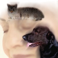
Typically involving neutral or pleasant emotional reactions.16,21,23 Any type of image can be seen, but the most common one is that of a human.12 Some patients report different hallucinations with each episode, while others see the same image each time.7 Patients usually do not recognize the people in their visions; although some will perceive images of themselves, possibly at an earlier stage in their lives. This type of imagery is known as heautoscopia.2
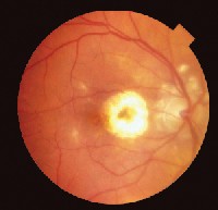 |
| Dry atrophic macular degeneration in an 84 year-old woman diagnosed with CBS. |
Also, the patient should not experience other delusions or hallucinations of the other senses.16,21,23 Interestingly, elderly patients who develop hearing loss may exhibit a similar phenomenon to CBS known as musical hallucinations, but this type of hallucination is much more rare.25
Loss of vision due to ocular disease is a specifying factor in most cases of CBS, but it is not obligatory for diagnosis.5 Because auditory hallucinations can occur, the coexistence of hallucinations of other modalities does not preclude a diagnosis of CBS, provided subjects are aware that their experiences are unreal.26
Management
The most critical management strategy for CBS is reassurance and counseling patients and their families or caregivers. Once patients understand that they are not crazy or going insane, the hallucinations usually do not bother them anymore. Patients more readily accept the hallucinations, and some patients even report enjoying these visions.14
For most patients, the visual hallucinations will likely disappear spontaneously, either with improvement or further deterioration of visual function.27 Even among patients who have no further vision change, hallucinatory episodes tend to occur less frequently over time.16
Pharmacotherapy has not been shown to be especially effective for patients who find the hallucinations annoying, upsetting or distracting.2 However, the anticonvulsants carbamazepine, clonazepam and valproate sodiumalong with the neuroleptics (antipsychotic drugs) thioridazine hydrochloride and haloperidolhave been used with some success.2
Moving patients into a more stimulating environment, keeping them socially involved and busy with hobbies and activities also can lessen the hallucinations.14,28 Surgical procedures that maximize the patients visual acuity (e.g., cataract surgery), meticulous refraction or low-vision aids can also be helpful.2
By educating ourselves about the history, pathogenesis, diagnosis and management of CBS, we can educate those who may have CBS or are at risk of developing it. And, we can ensure that patients will confide in us rather than hide in fear.
Dr. Skorin is the staff osteopathic ophthalmologist at Albert Lea Eye ClinicMayo Health System, Albert Lea, Minn.
1. Goldberg KB, Goldberg RE. Is seeing believing? Visual hallucinations in age-related macular degeneration and Charles Bonnet. J Ophthalmic Nurs Technol 2000 Jan-Feb;19(1):39-42.
2. Menon GJ, Rahman I, Menon SJ, Dutton GN. Complex visual hallucinations in the visually impaired. The Charles Bonnet syndrome. Surv Ophthalmol 2003 Jan-Feb;48(1):58-72.
3. de Morsier G. Les hallucinations. Rev Otoneuroophthalmol 1938;16:244-352.
4. Fenelon G. Visual hallucinations: the Charles Bonnet syndrome. Psychol Neuropsychiatr Vieil 2003 Jun;1(2):121-7.
5. Podoll K, Osterheider M, Noth J. The Charles Bonnet syndrome. Fortschr Neurol Psychiatr 1989 Feb;57(2):43-60.
6. Skorin Jr. L, Westberg M. Charles Bonnet syndrome: visual perception as deception. Clin Surg Ophthalmol 2004 Sep;22(9):242-52.
7. Teunisse RJ, Cruysberg JR, Hoefnagels WH, et al. Visual hallucinations in psychologically normal people: Charles Bonnet syndrome. Lancet 1996 Mar 23;347(9004):794-7.
8. Ball C. Charles Bonnet syndrome. Br J Psychiatry 1995 May;166(5):677-8.
9. Damas-Mora J, Skelton-Robinson M, Jenner FA. The Charles Bonnet Syndrome in perspective. Psychol Med 1982 May;12(2):251-61.
10. Fluornoy T. Le cas de Charles Bonnet. Arch Psych Swisse Romande 1902;1:1-23.
11. Fernandez A, Lichtshein G, Vieweg WV. The Charles Bonnet syndrome: a review. J Nerv Ment Dis 1997 Mar;185(3):195-200.
12. Burde RM, Savino PJ, Trobe JD. Clinical Decisions in Neuro-Ophthalmology. 3rd ed, St. Louis: C.V. Mosby, 2002:114-35.
13. Manford M, Andermann F. Complex visual hallucinations. Clinical and neurobiological insights. Brain 1998 Oct;121(Pt 10):1819-40.
14. Miller NR, Newmann NJ. Walsh & Hoyts Clinical Neuro-Ophthalmology: The Essentials, 5th ed. Baltimore: Williams & Wilkins, 1998;369-408.
15. Lessell S, Lessell IM, Glaser JS. Topical diagnosis: Retrochiasmal visual pathways and higher cortical function. In: Glaser JS (ed). Neuro-ophthalmology. 2nd ed. Philadelphia: J.B. Lippincott Co., 1990:213-38.
16. Schultz G, Melzack R. The Charles Bonnet syndrome: phantom visual images. Perception 1991;20(6):809-25.
17. Cohn R. Phantom vision. Arch Neurol 1971 Nov;25(5):468-71.
18. Gittinger JW Jr, Miller NR, Keltner JL, Burde RM. Sugarplum fairies. Visual hallucinations. Surv Ophthalmol 1982 Jul-Aug;27(1):42-8.
19. Teunisse RJ, Cruysberg JR, Verbeek A, Zitman FG. The Charles Bonnet syndrome: a large prospective study in The Netherlands. A study of the prevalence of the Charles Bonnet syndrome and associated factors in 500 patients attending the University Department of Ophthalmology at Nijmegen. Br J Psychiatry 1995 Feb;166(2):254-7.
20. Holroyd S, Rabin P, Finkelstein D, et al. Visual hallucinations in patients with macular degeneration. Am J Psychiatry 1992 Dec;149(12):1701-6.
21. Eagan SM, Williams JA. The formed visual hallucinations associated with vision loss. Optometry 2000 Aug;71(8):519-27.
22. Skorin Jr. L. Visual hallucinations. In: Onofrey BE, Skorin L, Holdeman NR (eds). Ocular Therapeutics Handbook: A Clinical Manual. 2nd ed, Philadelphia: Lippincott Williams & Wilkins, 2005:608-10.
23. Gold K, Rabins PV. Isolated visual hallucinations and the Charles Bonnet syndrome: a review of the literature and presentation of six cases. Compr Psychiatry 1989 Jan-Feb;30(1):90-8.
24. Schultz G, Needham W, Taylor R, et al. Properties of complex hallucinations associated with deficits in vision. Perception 1996;25(6):715-26.
25. Keating HJ. Music that hath no charms. Cortlandt Forum 2002 Aug; 15:30-2.
26. Hori H, Terao T, Nakamura J. Charles Bonnet Syndrome with auditory hallucinations: a diagnostic dilemma. Psychopathology 2001 May-Jun;34(3):164-6.
27. Tueth MJ, Cheong JA, Samander J. The Charles Bonnet syndrome: a type of organic visual hallucinosis. J Geriatr Psychiatry Neurol 1995 Jan;8(1):1-3.
28. Thorpe L. The treatment of psychiatric disorders in late life. Can J Psychiatry 1997;42(Suppl 1):19S-27S.
29. Lessell S. Visual hallucinations and related phenomena. Neur Neurosurg 1979;2:48-53.
30. Bracha HS, Wolkowitz OM, Lohr JB, et al. High prevalence of visual hallucinations in research subjects with chronic schizophrenia. Am J Psychiatry 1989 Apr;146(4):526-8.
31. Skorin L Jr. Visual hallucinations: perception as deception. Rev Optom 1993 Dec 15;130(12):30-2.
32. Bartlett JD. Ophthalmic toxicity by systemic drugs. In: Chiou GCY (ed). Ophthalmic Toxicology. 2nd ed. Philadelphia: Taylor & Francis, 1999:225-83.
33. Vaphiades MS, Celesia GG, Brigell MG. Positive spontaneous visual phenomena limited to the hemianopic field in lesions of central visual pathways. Neurology 1996 Aug;47(2):408-17.
34. Borruat FX. Visual hallucinations and illusions, symptoms frequently misdiagnosed by the practitioner. Klin Monatsbl Augenheilkd 1999 May;214(5):324-7.
35. Adamczyk DT. Am I seeing things? Optom Today 1999 June;39:37-9.
