You don't have to look to "a galaxy far, far away" to find optometrists manning the battle stations with lasers. Due to recent optometric scope expansions in Louisiana and Kentucky, added to the prior-existing scope-of-practice law in Oklahoma, clinicians using neodymium:yttrium-aluminum-garnet (Nd:YAG) lasers (1064 nanometers) to treat anatomically narrow angles can be found right here in the Milky Way.
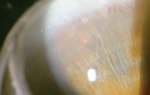 |
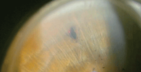
| In this right eye, at approximately 11 o’clock the clinician aims the laser directly to the left of the visible vessel to assure the laser doesn’t cause bleeding. Click image to enlarge. |
For new graduates, recently trained optometric residents and seasoned clinicians alike, the opportunity to treat anatomically narrow angles or angle closure using Nd:YAG lasers is growing. This article provides a step-by-step account of how to perform this procedure when the laws in your state permit.
The Four Angle Closures
The first step in disease management is a proper diagnosis. Taking careful steps at the beginning to fully understand the disease etiology and the orientation of the structures will make for a more predictable and positive outcome. Anatomically narrow angles can be sub-divided into four basic forms:
1. Pupillary block
2. Plateau iris configuration
3. Phacomorphic glaucoma
4. Malignant glaucoma
The end result of each abnormality is approximately the same; a blocked or narrow angle impeding the flow of aqueous humor entering and exiting the anterior chamber and increasing the intraocular pressure (IOP) by varying and, often, significant amounts. The appropriate diagnosis, however, is a critical element to appropriate treatment.
Pupillary Block
The most common form of angle closure, and the type most commonly associated with positive outcomes, is attributed to pupillary block.1 A pupillary block occurs when the pupil border comes in contact with the lens and shuts down the flow of the aqueous humor into the anterior chamber. The resultant iris bombé often forms, closing the angle and elevating the IOP. The application of a laser peripheral iridotomy (laser PI) is an immediate need in these cases and a successful procedure will typically lower the pressure within minutes after the procedure. The fellow eye should be carefully evaluated in this case as its angle formation may be similar. The opacification of the lens and anatomical structure, however, may be quite asymmetrical, placing one eye in jeopardy of a closed angle and not the fellow eye.
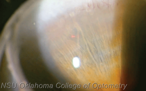 |
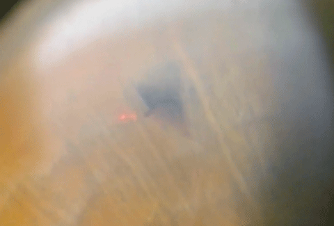
| Above, the clinician prepares to fire the first shot. Before beginning the procedure, we put Alphagan (brimonidine tartrate, Allergan) and pilocarpine in the eye. Below, after two shots, the impact of the laser is clearly visible as is much of the fluid rushing through the hole into the anterior chamber and helping to open up the patient’s angle. Click images to enlarge. |
 |
In many cases, the laser peripheral iridotomy (PI) is performed as a preventative procedure to prevent an acute closed angle.2 The old adage "an ounce of prevention is worth a pound of cure" is certainly true for this condition. If the standard Van Herrick angle estimation indicates a narrow angle, it is prudent to perform gonioscopy to visualize the angle and make a clinical judgment regarding the necessity of a PI. Anterior segment imaging is also particularly helpful in making a determination regarding the necessity of a preventative PI. Typically an angle less than 10 degrees is judged to be narrow and an indication to proceed with a PI. An angle less than 10 degrees as determined by anterior segment imaging typically corresponds to an angle in which the posterior pigmented trabecular meshwork cannot be observed by standard gonioscopy techniques. This gonioscopic view in which the posterior trabecular meshwork is not visible should be present in at least two quadrants before elevating the case to high closure risk and indicating the need for preventative PI.3 Another way of stating this is that if you can see the entire trabecular meshwork in three to four quadrants, then a laser PI is likely not indicated.
Plateau Iris Configuration
Another, much less common, angle closure occurs when the insertion of the iris root is displaced anteriorly. This is called plateau iris configuration. Due to the location of the iris root, a fold is created in the angle and bunched tissue places the angle at risk for closure. In previously undiagnosed cases, the patient may present with alarmingly elevated IOP (in the range of 40mm to 60mm) due to closed angles. A laser PI is usually performed within one to three days of closure onset, depending on the severity of the case and its initial response to medical management. Topical beta-blockers in one dose and brimonidine in one dose accompanied by topical steroids may be used as initial medical management. Carbonic anhydrase inhibitors are indicated if an IOP decrease is urgent. Pilocarpine should be used only in cases of phakic pupillary block or angle crowding.5
 |
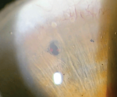
| More than two shots will be required, as a hole that is too small will heal easily and reclose the angle. The hole should be approximately 500µm, or about a 0.5mm. Click image to enlarge. |
It is prudent to treat the other eye prophylactically, assuming the patient responds well to the initial PI. If angle closure continues to be present after a patent PI the practitioner must have an elevated concern about plateau iris syndrome as opposed to plateau iris configuration. A PI will typically cure angle closure attacks in plateau iris configuration. In plateau iris syndrome, the peripheral iris bunches up in the angle and obstructs outflow without evidence of pupillary block. Often the diagnosis of plateau iris syndrome cannot be made until after evaluating the benefit of a PI. When treating suspected plateau iris configuration patients, be mindful that if the angle remains obstructed or recloses, the diagnosis of plateau iris syndrome must be carefully considered. Continued angle closure episodes even after a patent PI is a strong indication of plateau iris syndrome and a laser iridoplasty is the next clinical step to break the attack and prevent subsequent ones.
Phacomorphic Glaucoma
A rare form of angle narrowing or closure that occurs when the lens continues to pathologically enlarge and pushes the iris forward, narrowing the angle, is called phacomorphic glaucoma. A PI remains a strong component of early management but ultimately cataract surgery will be required.6
Procedural Checklist
|
Malignant Glaucoma
An extremely rare malady, malignant glaucoma, occurs when a misdirection of the flow of aqueous actually rotates in a cyclic pattern and pushes the vitreous forward, narrowing the angle. Typically this occurs after intraocular surgery in eyes with small anterior segments.7
Diagnosis
Good gonioscopy skills are necessary to visualize and appropriately evaluate the angle. Evaluate all 360 degrees of the angle. If the posterior trabecular meshwork is the most posterior structure visible, the angle can be assumed to be between 10 degrees and 20 degrees and is described as narrow—putting the patient at risk for angle closure. Clinical judgment and correlation with other symptoms and risk factors will be critical to advising these patients on the necessity of a PI. If only the anterior portion of the trabecular meshwork is visible, the angle is typically 10 degrees or smaller and the risk of closure is probable.8 This is the time to make recommendations to proceed with the PI. It is also relevant to evaluate pigment in the trabecular meshwork and check for neovascularization in the angle, as well as the presence of anterior synechiae. Prolonged, recurrent or subacute closed angle attacks that have occurred previously may be evident by residual peripheral anterior synechiae.
Thoroughly evaluate for contraindications before proceeding with PI treatment. Carefully consider corneal opacifications and abrasions. Ocular lasers typically cause intraocular inflammation, so be certain the eye is free from inflammatory disorders. The patient must be physically able to hold a steady fixation throughout the procedure. Although the patient cannot be dilated the day of the procedure, it is vital to assess the macula and retina prior to proceeding.
A risk-vs.-benefit dilemma is created when a narrow angle is identified in the initial slit lamp examination. Assessing the macula and retina is critical in most cases prior to proceeding with the procedure, but the question must arise: what if the dilation procedure creates the angle occlusion? If the patient presents with a closed angle, the standard of care and risk-vs.-benefit protocol would be to manage the most obvious and visually destructive condition, the closed angle. If the cornea is too cloudy to focus the laser beam, medical steps would need to be taken to reduce corneal edema. In severe cases, proceeding through a hazed cornea may be in the patient's best interest if medical management will not quickly decrease the pressure and edema. When addressing prophylactic concerns, dilate one eye at a time under carefully monitored conditions to assess the retina's and macula's condition prior to proceeding with the PI procedure. Monitor the patient carefully using only a minimal dilating agent, such as phenylephrine, and be certain a immediate danger has passed before scheduling the laser procedure.
Inform the patient that non-perforation or re-closure of the opening with time is a possibility. IOP spikes and inflammation are common and will be medically managed during the post-op period. Approximately 10% of patients undergoing the procedure will experience post-op pressure spikes.9 Also, inform the patient that, although unlikely, other risks include hyphema, synechiae, peaked pupil, floaters, monocular diplopia, retinal detachment and permanent vision loss.
After education and a signed consent form, pilocarpine is instilled prior to the procedure in an effort to constrict and tighten the iris for easy penetration. Typically, Alphagan or iopidine is instilled to retard pressure spikes. The decision to do both eyes at the same encounter is a personal one and calls for careful judgment. In many cases, it is in the patient's best interest to do both eyes at the same setting. Barring any complications, this is more efficient both in terms of time and monetary expense. The patient's health and visual welfare is always foremost and it is often prudent to do one eye and gauge the results in terms of complications, energy required and desired outcome before proceeding to the second eye.
 |
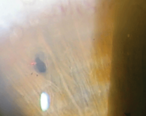
| Here, the clinician fires several more shots, allowing more pigment to come through the hole, equalizing the pressure between the posterior chamber and the anterior chamber. Click image to enlarge. |
Procedure Steps
After proparacaine is instilled in both eyes, both the patient and clinician should be seated, and adjustments should be made for comfort and decreased movement. Historically, the PI location is typically at 11 o'clock or 1 o'clock and should be tucked successfully under the upper lid. Although the PI location is still based primarily on personal preference and experience, an increasing amount of surgeons are placing the opening on the temporal meridian in an effort to reduce visual side effects such as dysphotopsia.10 It is best to identify a crypt or thinner area of the iris as the target.
Place the iridotomy lens on the anesthetized eye, cushioned with Celluvisc (carboxymethylcellulose sodium, Allergan) or a similar agent. Location of the 10mm 66D magnification insert section of the iridotomy lens is critical and should be aligned with the targeted area of the iris. After selecting the target, the optometrist's goal is to create an opening approximately 0.5mm to 1mm in diameter in a peripheral location that will not create a visual disturbance. The optometrist will observe a plume when penetration of the iris has occurred. The fluid behind the iris will rush forward through the opening, looking a bit like a cloud. Although this indicates penetration and can be dramatic, it is not the desired endpoint. Several additional shots will usually be necessary to widen the opening and prevent reclosure. The actual endpoint will come with experience and clinical judgment. The important aspect is to visualize the initial plume. The edges should be widened to 0.5mm to 1mm in diameter. Removing central strands will also prevent collection of debris in the center of the opening that may serve to eventually clog the created opening.
The YAG laser has a 95% penetration rate, making it the laser of choice. The spot size and duration are fixed. The energy necessary will vary tremendously based on iris thickness and pigmentation. Lighter, thinner irises are easier to penetrate. The numerical millijoules (mJ) value is typically between 2.0mJ and 5.0mJ. No focus offset is needed. Commonly the pulse is set on two. This means that with each fire of the trigger, two bursts of energy are delivered in rapid sequence. The power of each pulse of energy delivered is equivalent to the prescribed power set in millijoules. If 2.0mJ are set the laser will deliver 2.0mJ per pulse for a total energy power of 4.0mJ with the pulse counter set at two. The upper limit of total energy per eye applied in one setting is typically 150mJ, but can vary tremendously depending on clinical judgment and the energy necessary to create a patent opening.
The rule of thumb is to use the lowest energy and least number of shots to get the job done.
 |
| In the end, this patient’s angles went from only being able to see the anterior trabecular meshwork in two quadrants to being able to see scleral spur in all four quadrants, thanks to the procedure. Click image to enlarge. |
During the Procedure
There are a few caveats to be aware of that will make the experience more pleasant for both the doctor and the patient. A small amount of bleeding is quite common when a blood vessel is inadvertently struck by the laser. The blood will cascade momentarily, like a red waterfall, but with slight pressure applied to the eye with the iridotomy lens, the bleeding will typical cease and the procedure can be continued. On some occasions the doctor may have to pause momentarily to allow the field to clear.
The field can also be clouded by pigmentation, but this should not be confused with the plume described earlier. Although 150mJ of energy is considered the upper level, in a quiet eye nearing completion, it is often more prudent to finish the procedure with a few additional shots rather than have the patient return. Total energy in the eye should always try to be minimized as that reduces potential complications. Clinical experience has shown energy levels between 15mJ and 200mJ to be optimal for most laser PIs, with lighter colored blue eyes often having thinner irides and requiring much less energy.
Always try to eliminate strands of tissue crisscrossing the opening, as this will often clog the opening with time and decrease the effectiveness. Patients will often feel slight pain if the per-shot energy level is higher than their tolerance threshold. Patients should be instructed to report pain or uncomfortable sensations during the procedure so that the energy can be decreased accordingly in small increments.
Postoperative Care
After successfully creating a patent opening, one drop of Alphagan or iopidine should be instilled post-operatively. The patient should remain in the clinic for approximately 30 minutes to evaluate for postoperative pressure spikes. If the pressure is rising, instill more pressure reducing medication and keep the patient in the office until the pressure is under control. Upon dismissal the patient should be placed on prednisolone acetate four times daily for one week followed by a rapid taper to decrease the inflammatory response. Alphagan is typically not needed after the first hour, but if the pressure is still of concern Alphagan can be used twice a day until the return visit one week later. In cases when Alphagan is contraindicated, consider timolol or oral acetazolamide.
May the Force be with Your Patients
As with any procedure, education can help alleviate the patient's concerns. Educate the patient of the possible common side effects, such as IOP elevations and inflammation, and how these will be addressed. Also, review the rare complications, such as retinal detachment, and the signs and symptoms to watch for. After reviewing any concerns the patient may have and addressing those, evaluation at the one-week visit should include visual acuities, IOPs, slit lamp evaluation, gonioscopy and retinal inspection. Anterior segment imaging and quantifying the angle will also assist in determining the overall benefit of the procedure. If no complications are identified at the one-week follow-up, the patient can then be returned back to primary eye care.
Recognizing patients who would benefit from this procedure, providing quality education and preventing permanent sight loss is a win/win in any galaxy.
Dr. Miller is a professor of optometry at the NSU Oklahoma College of Optometry where he oversees the glaucoma clinics and course.
Dr. Shetler is an assistant professor and chief of the university clinic facilities at the Oklahoma College of Optometry.
Dr. Lighthizer is the assistant dean for clinical care services, director of continuing education, and chief of both the specialty care clinic and the electrodiagnostics clinic at NSU Oklahoma College of Optometry.
|
1. Allingham R, Damji K, Freedman S, et al. Shields Textbook of Glaucoma 6th ed. Philadelphia: Lippincott, 2011. 2. Al-Aswad L, Tsai J. Managing the Narrow-angle patient. Review of Ophthalmology. 2004;4(6):45-50. 3. Foster P,Buhrmann R, Quigley HA, Johnson GJ. The definition and classification of glaucoma in prevalence surveys. Br J Ophthalmology 2002;86:238-42. 4. Allingham R, Damji K, Freedman S, et al. Shields Textbook of Glaucoma 6th ed. Philadelphia:Lippincott, 2011. 5. Wills Eye Hospital, The Wills Eye Manual 3rd ed. Lippincott Williams & Wilkins,1999. 10;10 :252-3. 6. Tomey K, al-Rajhi A. Neodymium:YAG laser iridotomy in the initial management of phacomorphic glaucoma. Ophthalmology. 1992;99(5):660–5. 7. Allingham R, Damji K, Freedman S, et al. Shields Textbook of Glaucoma 6th ed. Philadelphia: Lippincott, 2011. 8. Foster P, Buhrmann R, Quigley H, Johnson G. The definition and classification of glaucoma in prevalence surveys. Br J Ophthalmol. 2002;86:238-42. 9. Jiang Y, Chang D, Foster P, et al. Immediate changes in intraocular pressure after laser peripheral iridotomy in primary angle-closure suspects. Ophthalmology. 2013;119(2):283-8. 10. Vera V, Naqi A, Belovay G, et aI. Dysphotopsia after temporal versus superior laser peripheral iridotomy: a prospective randomized paired eye trial. Am J Ophthalmol. 2014; 157(5):929-35. |
