 |
History
A 53-year-old white male presented for a routine eye examination without visual or ocular complaints.
His ocular history was remarkable for what he called a birth mark in the back of his right eye. His systemic history was noncontributory.
Diagnostic Data
His best-corrected acuity was 20/20 O.U. at distance and near. External examination was normal, and there was no evidence of afferent pupillary defect.
Biomicroscopy demonstrated normal anterior segment structures and open angles. Applanation intraocular pressure measured 20mm Hg in both eyes.
The pertinent fundus finding is demonstrated in the photograph.
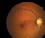 |
| The patients retina O.D. on presentation. |
Your Diagnosis
How would you approach this case? Does this patient require additional tests? What is your diagnosis?
How would you manage this patient? What is the likely prognosis?
Discussion
The diagnosis in this case is magnocellular nevus, otherwise known as melanocytoma. Melanocytomas are benign, deeply pigmented tumors that arise from dendritic melanocytes incorporated within the optic disc. As an entity, they are more common in blacks and Mediterranean races.1,2
Melanocytomas may arise in any part of the uveal tract. Commonly, they are situated eccentrically on the disc and project 2mm or less into the vitreous. They may extend into the lower temporal retina posteriorly, beyond the lamina. The surface of the lesion commonly appears striated because of the interdigitation of darkly pigmented tumor cells between axons in the nerve fiber layer. If these lesions compress the optic disc, prominent visual field defects are possible. These lesions may enlarge, but growth is almost always slow.1,2
Melanocytomas should be differentiated from other tumors of the disc, such as astrocytic hamartoma, which may be seen with tuberous sclerosis or neurofibromatosis, rare pigment epithelial proliferations, hemangioma and peripapillary choroidal melanoma.1,2
While melanocytomas do not metastasize, they must still be distinguished from peripapillary uveal melanoma of the optic nerve. The borders of a melanocytoma, as seen by ophthalmoscopy, tend to feather out, in contrast to the borders of a melanoma.1,2
Additional diagnostic testing may include observation using red-free lighting, neuroimaging, B-scan ultrasound, fluorescein angiography, fine needle aspiration biopsy and photodocumentation.1,2
1. Gurwood AS, Pagani JM, Eagan R, Lee S. Rare dual finding: Melanocytoma and optic pit. Review of Optometry 1993; 130(9):79-82.
2. Alexander LJ. Congenital and Acquired Anomalies of the Optic Nerve Head. In: Alexander LJ. Primary Care of the Posterior Segment, 2nd ed. Norwalk, CT: Appleton and Lange; 1994:89-170.
