 |
Q:
I recently saw a patient with a mature hand-motion cataract who also had an obvious afferent pupillary defect (APD) in that same eye. Is the cataract the cause?A:
“It is important to understand that a cataract, no matter how asymmetric in presentation, will never cause a relative afferent pupillary defect in that eye,” says Kelly Herron, OD, of Omni Eye Services in Atlanta. “It may seem counterintuitive, but instead of acting like a filter, a cataract can actually increase the light stimulus compared with the other eye.”
If there is an APD present, even in the setting of a mature, unilateral cataract, another etiology must be suspected and investigated. One can detect an APD when there is asymmetric damage to the afferent visual system. This damage can occur anywhere from the retina to the anterior part of the optic tract. Considering the seriousness of the pathology usually responsible for an APD, it is essential to check pupils thoroughly at every exam and explain an APD, if present.
When checking for an APD, use a bright light and hold it close enough to isolate the pupillary response for each eye. Dr. Herron recommends a binocular indirect ophthalmoscope as a reliably bright source. An APD that is apparent immediately with the swinging flashlight test is significant and usually obvious to the clinician. To catch smaller APDs, Dr. Herron advises performing the “three-second swinging flashlight test,” where the light source is held for three seconds in front of each pupil to watch for a quicker release in one of the pupils.
Also, have the patient fixate at a distance target to confirm that the convergence/accommodative response is not in play. If there is a question whether an APD is present, perform supportive subjective tests, such as light brightness comparison between eyes. Unilateral abnormalities found in any test will also point toward a diagnosis of an APD.
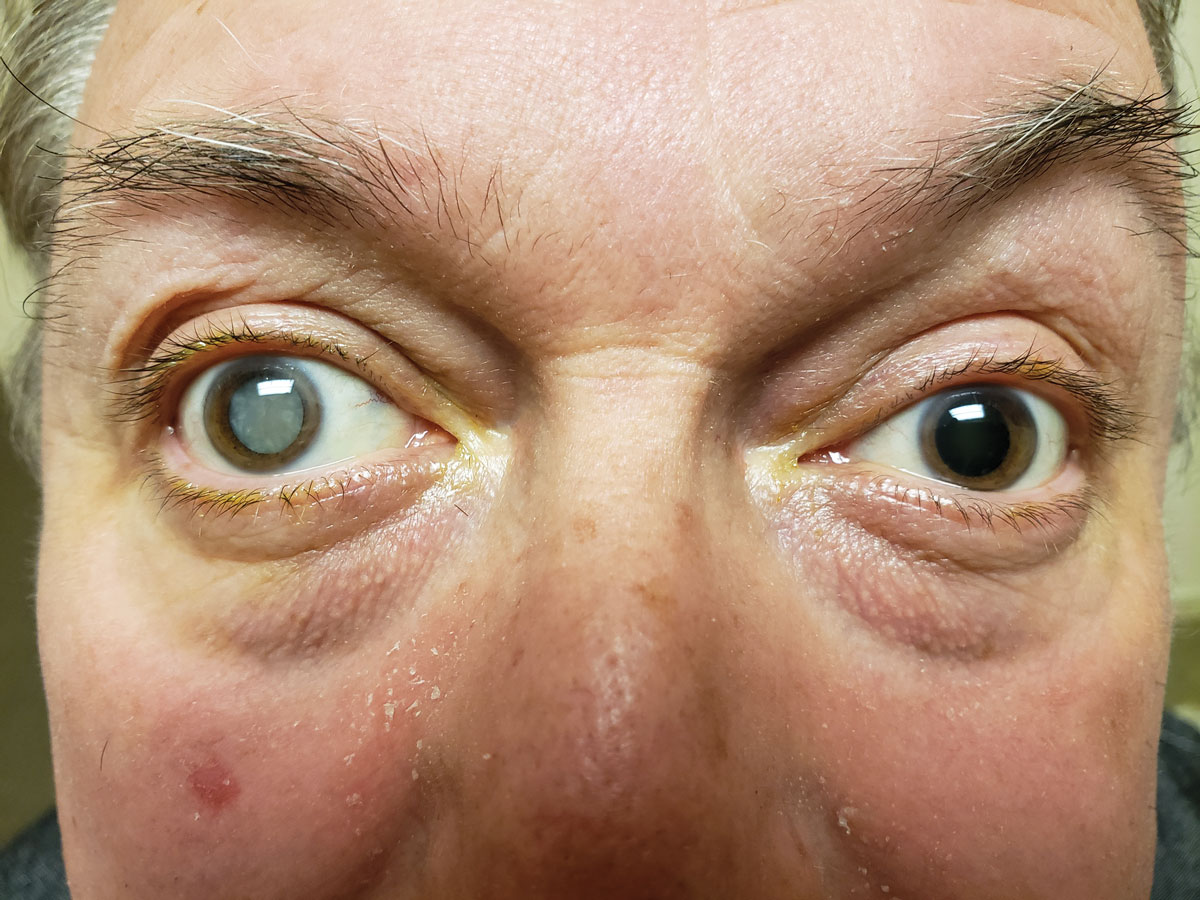 |
| Was a dense cataract the cause of this patient’s APD? Click image to enlarge. |
In-depth History
Once the diagnosis of an APD is made, a thorough chief complaint and history should guide the rest of your exam and workup. Pathology responsible for causing an APD can include significant amblyopia, major retinal issues and optic neuropathy. “I typically take a systematic approach and start with the patient’s current symptoms and then move on to the systemic and ocular history,” Dr. Herron says.
“Since this patient’s visual symptoms are at least in part due to the cataract, we chose to focus on other associated issues,” Dr. Herron notes. After basic glaucoma history questions, she explored neurological and giant cell arteritis possibilities.
The answer was not clear until Dr. Herron asked her patient about any history of eye/head trauma or poor vision/patching as a child. At first, the patient denied any injury, but upon further questioning, he revealed that he remembered being in a car accident years ago. This prompted the team to continue their exam with tonometry, gonioscopy and a dilated fundus exam that revealed increased IOP, angle recession and a traumatic cataract in the right eye only.
“Our diagnosis of an APD in the setting of angle recession glaucoma made sense with our clinical findings and lack of any other concerning symptoms or health history,” Dr. Herron says. “The cataract was removed, revealing a significantly cupped optic nerve consistent with glaucoma. If the etiology remained unclear, a systemic or neurology workup would have been indicated.
“Checking pupils is essential to every exam and can reveal many important findings about the eyes and brain,” Dr. Herron says. “Investigate and explain an APD to ensure your clinical findings make sense and the patient gets the proper workup and care.”
