Throughout the year, nearly all optometrists see a plethora of patients who present with signs and symptoms of allergic eye disease. Clearly, the same can be said of allergists and immunologists.
Caring for patients who present with allergic eye disease represents a unique area of opportunity between eye care professionals and allergists. Drawing upon clinical experience, cross-specialty referrals unquestionably facilitate more accurate diagnoses and improved patient outcomes.
This article reviews the signs and symptoms of allergic eye disease, with specific clinical insight. Additionally, it discusses the most effective testing options and treatment strategies for your ocular allergy patients.
Immunology of the Anterior Surface
Allergic
manifestations of the ocular surface have been observed in patients
who’ve undergone LASIK surgery.12 Typically, allergy-related
complications manifest in those with increased postoperative irritation,
visual morbidity and dry eye complications.
LASIK
causes significant inflammation in normal patients, which is further
exacerbated in atopic eyes. Further, a diffuse lamellar keratitis is one
particular complication of LASIK that has been linked to atopic
individuals.12 In general,
goblet cell density decreases in patients with allergic inflammation
and/or dry eye disease. Additionally, LASIK has been associated with
decreased goblet cell density and mucin volume.13,14 Therefore,
patients with pre-existing allergic conjunctivitis and dry eye disease
should be managed appropriately during the preoperative evaluation
process for LASIK surgery.
The eye is an immunologically distinct entity—it has a unique anterior chamber immune system; it lacks formed lymph nodes within the orbit, lacrimal, gland, eyelids and conjunctiva; and it contains one of the densest populations of mast cells (approximately 50 million) located beneath the conjunctival surface and the pericular tissue.
LASIK’s Impact on Allergy
The conjunctiva is composed of thin, non-keratinizing squamous epithelial cells with varying densities of goblet cells, Langerhan’s cells and stem cells––all of which are predominantly found at the limbus. The conjunctiva extends from the lid margin to the limbus. Thus, conjunctival inflammation can be triggered not only by allergens that penetrate the epithelial surface into the substantia propria (where the mast cells reside), but also by contact dermatitis, dermatophyte infestation or stem cell damage following LASIK surgery (see “LASIK’s Impact On Allergy.”).
The anterior surface is bathed in a tear biofilm that exhibits a lipid, aqueous and mucoid layer. Additionally, the tears contain a host of biologically active mediators (e.g., immunoglobulins, cytokines, interleukins, growth factors) that alter the character of the tear biofilm, based on the underlying inflammatory response of the ocular surface.
Pathophysiology of AC
Allergic conjunctivitis (AC) triggered by IgE-mast cell-mediated activation is the most common hypersensitivity of the ocular surface. It is generally understood that the condition affects up to 40% of the American population.
Direct exposure of the ocular mucosal surface to the patient’s surrounding environment activates mast cells. This process occurs when an allergen attaches to its specific epitopes via the IgE antibody and binds to the mast cell surface. Once two IgE molecules crosslink to the allergen, the mast cell is activated, which yields the acute and late phases of AC.
In the late phase of AC, the conjunctival mucosal surface is infiltrated by neutrophils, eosinophils, lymphocytes and macrophages. The presence of eosinophils in the conjunctival surface confirms the diagnosis of AC.
Signs and Symptoms of AC
Mediators of IgE-Mast Cell Activation in Allergic Conjunctivitis15,16
The signs and symptoms of ocular allergy are casued by mast cell activation via specific allergen-crosslinking IgE molecules. IgE-mast cell activation permits the immediate release of preformed histamine, which yields the early-phase allergic reaction; activates various enzymes that assist in the formation of leukotrienes and prostanglandins; and releases multiple cytokines, which initiates the late-phase reponse.
Preformed allergies
De novo allergies
The allergic inflammatory reponse involves the four classic signs of inflammation: rubor (redness), tumor (swelling), calor (warmth) and dolor (pain).
The mixture of preformed and de novo mediators provides various opportunities for a synergism between the signs and symptoms of allergy (see “Mediators of IgE-Mast Cell Activation in Allergic Conjunctivitis."). And it’s this specific constellation of signs and symptoms that helps guide your therapeutic mangement plan, including the prescription of agents that inhibit more than one allergic mediator.
A particular caveat for eye care specialists: “Ocular pain” is not suggestive of acute allergic inflammation. Instead, such a complaint should prompt you to investigate for either a chronic allergic condition, such as atopic keratoconjunctivitis (AKC), vernal keratoconjunctivitis (VKC), or other ocular conditions such as optic neuritis, uveitis, iritis, keratitis or scleritis. Blepharospasm can occur in patients with AKC or VKC, and both conditions often generate excessive, sticky mucus and photophobia upon morning waking.
Seasonal vs. Perennial AC
More than two decades ago, researchers believed that the most common form of ocular allergy was seasonal allergic conjunctivitis (SAC), while perennial allergic conjunctivitis (PAC) was reported to affect just 5% of the population.1 More recent reports based on the National Health and Nutrition Survey III suggested that ocular symptoms associated with allergies affect 40% of the US population, with an increased prevalence of perennial allergen-linked symptoms.2 The progressive prevalence of ocular allergy appears to be multifactorial, and has been linked to epigenetics; climate change; and a confounding increase in other anterior surface conditions, such as multiple forms of dry eye syndrome.2
The onset of SAC coincides with increased environmental pollen release. By age 30, more than 80% of patients who exhibited nasal allergies during preadolescence will experience SAC.3 Multiseasonal exacerbations during the tree, grass and weed seasons are common and may persist for as long as four weeks, depending on the pollen production cycle of the offending plant species.
PAC is commonly associated with perennial allergens, such as molds, dust mites and animal dander. Keep in mind that the pollination seasons of some plants and trees persist longer than four weeks. In such instances, the associated allergic conjunctivitis potentially could be considered perennial by some clinicians.
Allergy Testing
• Patient history. Obtaining a thorough patient history usually provides general direction when seeking a diagnosis of any allergic presentation. Further, a careful history helps uncover the presence of other comorbid conditions. However, the history alone may not provide enough information to determine which specific allergen is affecting the patient.4,5 In allergy management, the clinical diagnosis—as determined by the history and physical examination—is supported by an assessment of the IgE antibodies. The diagnosis of conjunctivitis or allergic rhinoconjunctivitis can be detected in more the sweeping majority of patients.
Remember that allergic disorders rarely affect a single organ. In most cases, allergic reactions include any combination of several primary symptoms, such as asthma, allergic rhinitis, rhino-conjunctivitis, atopic dermatitis or urticaria.
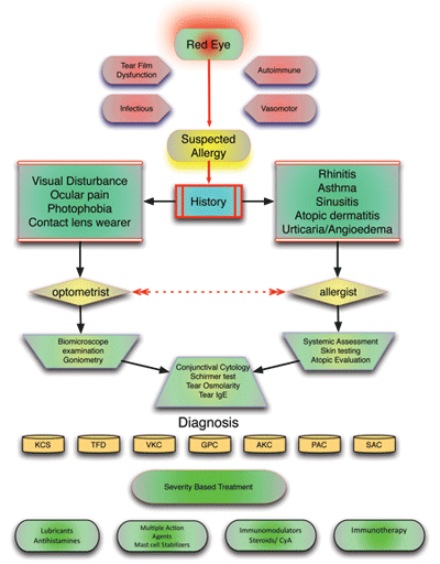
Proper evaluation of a red eye involves analyzing a wide spectrum of disorders. To treat these signs and symptoms of ocular allergy, patients should seek care from either an eye care provider or an allergist—the health care providers who are best suited to help minimize symptoms and optimize visual outcome.
• Physical examination. Begin the examination of an ocular allergy patient with a simple inspection of the face and area surrounding the eye. This includes an evaluation of the periocular tissue and eyelids for evidence of dermatitis, swelling, discoloration, ptosis or blepharospasm, as well as the presence or absence of tear discharge.
Additional clues for ocular allergy include the presence of a horizontal skin crease across the bridge of the nose (aka, “nasal salute”) that occurs in patients who constantly rub their nose due to itching. “Allergic shiners” are ecchymotic-looking areas located beneath the eyes typically are associated with allergic rhinitis. Periorbital edema is common, and is more likely to develop on the lower lids secondary to gravitation forces.4
Following facial inspection, examine the conjunctiva for signs of chemosis, hyperemia, cicatrization or papillae formation on the palpebral and bulbar membranes.
• Skin prick testing. Intradermal and skin prick tests are used to confirm a specific allergen sensitivity. Both diagnostic techniques are simple, rapid and inexpensive. Also, because the patient readily observes either process, the skin testing procedure may serve as part of the allergy education process.
Keep in mind, however, that the conjunctivae may be uniquely sensitized to a particular allergen. So skin testing often proves negative in these instances, and patients may subsequently require allergen-specific serum IgE testing or conjunctival provocation with specific allergens (performed at specialized centers) to further evaluate the localized conjunctival response.
• Patch testing. Patch testing is an essential investigative technique that helps the clinician identify specific allergens in cases of delayed-type hypersensitivity reaction, such as contact dermatitis.
This process involves the detection of primed, antigen-specific T-lymphocytes that circulate throughout the body using non-irritating antigen application to normal skin. Within two to three days after patch application, the individual’s skin is evaluated for allergic response.
Eye care providers, in particular, should be aware that some patients may test positive for allergies to benzalkonium chloride and thimerosal––preservative agents currently or historically found in many ophthalmic medications and contact lens solutions.6
• Serum IgE. Detection of allergen-specific IgE in the blood serum is used when skin or patch testing is ineffective or impractical. These instances include:
To maximize treatment efficacy, the clinician must be able to differentiate between the allergic and non-allergic inflammatory conditions. This includes a proper patient history with a detailed symptom list; a physical examination that includes disease signs; and the determination of a primary diagnosis.
- Poor patient cooperation during skin testing.
- Patients who have extensive dermatitis.
- Patients on long-term topical and/or systemic antihistamine therapy.
Additionally, allergen-specific IgE testing allows us to quantify sensitizations to diverse antigens simultaneously. Keep in mind, however, that skin testing has been shown to have a higher sensitivity and specificity than serum-based testing.
Ocular Allergy Treatments
Patients with severe ocular allergies may seek the immediate assistance of an optometrist to resolve the underlying allergic inflammation, ensure eye health and preserve vision. Many patients with allergic conjunctivitis also require a more extensive evaluation for comorbid atopic disorders.
Our armamentarium for ocular allergy includes:
• Nonpharmacological treatment. Nonpharmacological treatment consists of allergen avoidance, preservative-free ocular lubricants, cold compresses and the use of daily disposable contact lenses.
• Histamine H1 receptor antagonists. Antihistamines bind with histamine receptors, which decrease vasodilation and instantly relieve the primary symptom of itching. These agents have a minimal effect on conjunctival erythema. Oral antihistamines are used for mild allergies that involve nasal symptoms; however, many of these drugs have some anticholinergic effect and may augment the surface drying effect associated with ocular allergy.7 Common oral histamine antagonists include diphenhydramine, loratadine and fexofenadine.
• Mast cell stabilizers. Mast cell degranulation is the primary site of allergic inflammation. Mast cell stabilization prevents the release of proinflammatory allergic mediators. However, to be clinically maximizing their effectiveness, mast cell stabilizers must be taken prophylactically––before allergen exposure. Common mast cell stabilizers include cromolyn, lodoxamide, nedocromil and pemirolast.
• Topical mast cell stabilizers/H1 receptor antagonists. Newer ophthalmic allergy medications provide dual (and multiple) action by simultaneously inhibiting mast cell degranulation and preventing histamine release. Common topical mast cell stabilizers/H1 receptor antagonists include alcaftadine, azelastine, bepotastine, epinastine and olopatadine.
• Topical non-steroidal anti-inflammatory drugs (NSAIDs). Topical NSAIDs inhibit prostaglandin synthesis via blockage of cyclooxygenase (COX1 and COX2)––the enzyme responsible for the production of inflammatory mediators. COX inhibition reduces tissue inflammation and alleviates symptoms of ocular itch. Common examples of topical NSAIDs include ketorolac and bromfenac.
| Recommendations for Cross-Specialty Consultations |
|
Eye care providers should refer ocular allergy patients to an allergist when:
Allergists should refer ocular allergy patients to an eye care provider when:
|
• Allergen immunotherapy. High-dose, subcutaneous allergen immunotherapy is a well-established treatment for allergic conjunctivitis and rhinitis. Immunotherapy not only affords the individual increased tolerance to allergen exposure, but also decreases his or her dependence upon medication.
One study showed that, following immunotherapy, patients could tolerate exposure to significantly higher allergen concentrations before exhibiting symptoms of redness, pruritus and ocular swelling.10,11
Dr. Bielory is an attending at Robert Wood Johnson University Hospital and principal investigator, US EPA grant on Climate Change and Allergic Airway Disease, Rutgers University in New Brunswick, NJ.
1. Dart JK, Buckley RJ, Monnickendan M, Prasad J. Perennial allergic conjunctivitis: definition, clinical characteristics and prevalence. A comparison with seasonal allergic conjunctivitis. Trans Ophthalmol Soc U K. 1986;105 ( Pt 5):513-20.
2. Singh K, Axelrod S, Bielory L. The epidemiology of ocular and nasal allergy in the United States,1988-1994. J Allergy Clin Immunol. 2010 Oct;126(4):778-783.e6.
3. Rosario N, Bielory L. Epidemiology of allergic conjunctivitis. Curr Opin Allergy Clin Immunol. 2011 Oct;11(5):471-6.
4. Dinowitz M, Rescigno R, Bielory L. Ocular allergic diseases: differential diagnosis, examination techniques, and testing. Clin Allergy Immunol. 2000;15:127-50.
5. Williams PB, Siegel C, Portnoy J. Efficacy of a single diagnostic test for sensitization to common inhalant allergens. Ann Allergy Asthma Immunol. 2001 Feb;86(2):196-202.
6. Hong J, Bielory L. Allergy to ophthalmic preservatives. Curr Opin Allergy Clin Immunol. 2009 Oct;9(5):447-53.
7. Wade L, Bielory L, Rudner S. Ophthalmic antihistamines and H1-H4 receptors. Curr Opin Allergy Clin Immunol. 2012 Oct;12(5):510-6.
8. Bielory BP, Perez VL, Bielory L. Treatment of seasonal allergic conjunctivitis with ophthalmic corticosteroids: in search of the perfect ocular corticosteroids in the treatment of allergic conjunctivitis. Curr Opin Allergy Clin Immunol. 2010 Oct;10(5):469-77.
9. Bielory L. Ocular symptom reduction in patients with seasonal allergic rhinitis treated with the intranasal corticosteroid mometasone furoate. Ann Allergy Asthma Immunol. 2008 Mar;100(3):272-9
10. Bødtger U. Prognostic value of asymptomatic skin sensitization to aeroallergens. Curr Opin Allergy Clin Immunol. 2004 Feb;4(1):5-10.
11. Bødtger U, Poulsen LK, Jacobi HH, Malling HJ. The safety and efficacy of subcutaneous birch pollen immunotherapy. Allergy. 2002 Apr;57(4):297-305.
12. Bielory BP, O’Brien TP. Allergic complications with laser-assisted in-situ keratomileusis. Curr Opin Allergy Clin Immunol. 2011 Oct;11(5):483-91.
13. Albietz JM, Lenton LM, McLennan SG. Effect of laser in situ keratomileusis for hyperopia on tear film and ocular surface. J Refract Surg. 2002 Mar-Apr;18(2):113-23.
14. Rodriguez AE, Rodriguez-Prats JL, Hamdi IM, et al. Comparison of goblet cell density after femtosecond laser and mechanical microkeratome in LASIK. Invest Ophthalmol Vis Sci. 2007 Jun;48(6):2570-5.
15. Bielory L. Allergic and immunologic disorders of the eye. Part I: immunology of the eye. J Allergy Clin Immunol. 2000 Nov;106(5):805-16.
16. Bielory L. Ocular allergy overview. Immunol Allergy Clin North Am. 2008 Feb;28(1):1-23, v.
