 A 69-year-old black female presented for an ophthalmic evaluation at the request of her hematologist/oncologist. Her chief complaint was recent, progressive, bilateral visual blur at distance and near with her spectacles. Her systemic history was remarkable for hypertension, hypercholesterolemia and migraine headache. In addition, she was diagnosed with multiple myeloma one month prior.
Systemic medications included Zestril (lisinopril, AstraZeneca), Microzide (hydrochlorothiazide, Watson Laboratories), Lipitor (atorvastatin, Pfizer) and low-dose (81mg) aspirin. She was being monitored frequently by both her internist and hematologist/oncologist.
Diagnostic Data
Hyperviscosity in MM may cause a reduction of axoplasmic flow, resulting in cotton-wool infarct and nerve fiber dropout.
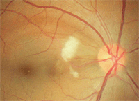
Her best-corrected distance acuity was 20/25 O.U. Pupils were equally reactive, with no afferent pupillary defect. Biomicroscopy revealed mild, bilateral nuclear and cortical lens opacities. Intraocular pressure measured 14mm Hg O.U. at 9:00a.m. Dilated funduscopy revealed two cotton-wool spots in the papillomacular region of the right eye. Nerve fiber layer dropout was seen superior-temporal to the disc in that eye, adjacent to the cotton-wool infarct (see figure).
Amsler grid testing of her right eye revealed a distorted area infero-nasal to fixation. Retinal blood vessels showed grade 2 arteriolar sclerosis in both eyes. Optic nerves appeared healthy, with a cup-to-disc ratio of 0.30 x 0.30 O.U. Her blood pressure measured 140/88mm Hg (right arm sitting).
We scheduled the patient to return for central threshold perimetry and nerve fiber layer analysis. We sent summary reports to the patient’s internist and oncologist.
The presence of cotton-wool infarct could be secondary to our patient’s hypertension. Could her multiple myeloma also be responsible for the retinal appearance?
What is Multiple Myeloma?
Multiple myeloma (MM) is a rare cancer characterized by the proliferation of malignant B cells (plasma cells) in the bone marrow and subsequent overabundance of antibodies.1-3 The monoclonal protein is IgG in 50% of cases, IgA in 25% of cases, and IgD or IgE much less commonly. Immunoglobulin G (IgG) is usually the elevated protein in MM.2,4,5
Despite recent therapeutic advances that have improved the quality of life and length of survival, MM is still incurable.1,5,6 Early diagnosis is difficult because symptoms may not appear until the cancer is advanced. The average five-year survival rate is about 30%.1-3
C = Calcium (elevated) R = Renal failure A = Anemia B = Bone lesions
Who Gets MM?
CRAB: Mnemonic for Complications of MM
In the United States, approximately 45,000 people are living with MM. It is the second most prevalent blood cancer after non-Hodgkin’s lymphoma. MM is more commonly diagnosed in persons over 65 years of age, and more common in men than in women, and in blacks than in whites.2,3 Age-adjusted annual incidence is 3 cases per 100,000 white women, 4.3 cases per 100,000 white men, 6.7 cases per 100,000 black women and 9.6 cases per 100,000 black men.1,5,6 Exposure to high amounts of radiation, herbicides, insecticides, petroleum products, heavy metals, plastics, and asbestos appear to be risk factors.6
Complications of MM
Although the tumor cells remain primarily within the bone marrow, MM may cause a wide variety of organ and organ system dysfunctions that may include ophthalmic involvement.1,2 The overproduction of plasma cell antibodies may lead to hyperviscosity, amyloidosis and renal dysfunction/failure.1,3 Generalized fatigue, weight loss and bone pain on movement are frequent symptoms.1,3
Hypercalcemia (30% of patients at presentation), susceptibility to infections (pneumonia, bladder or kidney infection, sinusitis), congestive heart failure, bleeding problems from thrombocytopenia, spinal cord compression and neurologic symptoms (weakness or numbness) can also occur.1,2,7,8 Hyperviscosity syndrome may develop secondary to the high volume of monoclonal protein. Stroke, myocardial ischemia and myocardial infarction also may result.5 Common causes of death in MM patients are infection, hematologic complications and renal failure.5
Differential Diagnosis of Multiple Myeloma • Monoclonal gammopathy of undetermined significance (MGUS) • Waldenström’s macroglobulinemia • Amyloidosis • Cancer deposits in bone due to spread from breast or lung cancer • Other blood malignancies, such as lymphoma or leukemia
The Eye in MM
Ocular signs of MM may appear at the initial presentation or develop later in the disease process. They result from myeloma infiltrates or the associated hematologic sequelae. In the retina, cotton-wool spots, nerve fiber dropout, hemorrhages and vascular abnormalities (including dilation, tortuosity and microaneurysms) can develop secondary to the associated elevated serum viscosity.
Optic nerve, oculomotor nerve and lacrimal gland infiltration by myeloma cells have been reported. Iridescent crystalline or copper deposits of the conjunctiva and cornea, ciliary body cysts and cranial nerve compression causing palsies have also been described in MM.2,3
Diagnosing MM
MM may be discovered by a primary care physician during a routine physical examination and by using blood and urine tests. Serum protein electrophoresis (SPEP) separates blood proteins and can detect the presence of M proteins (monoclonal immunoglobulin) called an “M spike.” When found in urine, the M proteins are referred to as Bence Jones proteins.7,9
A complete blood count (CBC) can determine if the patient has anemia, thrombocytopenia or leukopenia. A comprehensive metabolic panel, including blood urea nitrogen (BUN), creatinine and uric acid, also should be performed. SPEP, urine protein electrophoresis (UPEP) and immunofixation are used to determine the type and subtype of each protein present. Quantitative immunoglobulin (i.e., IgG) levels, beta-2 microglobulin, C-reactive protein and erythrocyte sedimentation rate (ESR) may also be performed.7
Imaging studies of suspected MM patients may include a skeletal series of X-rays of the skull, long bones (looking for impending fractures) and axial skeleton.9 MRI scans of the vertebrae are often diagnostic when plain radiographs are not. Therefore, symptomatic patients are evaluated with MRI to obtain a clear view of the spinal column and to assess the integrity of the spinal cord. CT scan is occasionally performed to measure the size of soft tissue plasmacytomas.7,9
Bone marrow biopsy is usually performed to calculate the percent of bone marrow occupied by plasma cells (infiltration). Biopsy is also helpful in identifying sheets or clusters of plasma cells in the specimen.
Treatment and Management
Treatment of MM is focused on disease containment and suppression.4 If patients are asymptomatic, with no end-organ damage, treatment may be deferred. In addition to drugs that inhibit the destruction of bone, conventional treatment for multiple myeloma involves chemotherapy and corticosteroids. Bone marrow transplantation also may be beneficial.9
Ongoing research aims to elucidate the etiology of MM and develop more effective treatments. Newer approaches include immunomodulatory therapy and targeted agents. Adjunctive therapy includes radiation to target areas of pain, impending pathologic fracture, or existing pathologic fracture.1,2,4,8,9
For our patient, we scheduled her to return in eight weeks to re-evaluate the cotton-wool infarct. But we could not determine whether its etiology is from her hypertension or from MM. Her hypertension remains stable, so we will continue to monitor her.
MM is a debilitating malignancy that results in a multi-system disease. The primary ocular manifestation is hyperviscosity-related retinopathy, although myriad ophthalmic sequelae can occur. Patients with MM require careful follow-up ophthalmic visits and comanagement with hematology/oncology to ensure optimal care. Awareness of the symptoms, signs and manifestations of MM may lead to an earlier diagnosis and have a positive influence on the disease course.
1. Bataille R, Harousseau JL Multiple myeloma. N Engl J Med. 1997 Jun 5;336(23):1657-64.
2. Myers H. Dysproteinemias, ch. 70. In: Primary Eyecare in Systemic Disease. Norwalk, CT: Appleton and Lange, 1995.
3. Fung S, Selva D, Leibovitch I, et al. Ophthalmic manifestations of multiple myeloma. Ophthalmologica. 2005 Jan-Feb;219(1):43-8.
4. Orellana J, Friedman AH. Ocular manifestations of multiple myeloma, Waldenstrom’s macroglobulinemia and benign monoclonal gammopathy. Surv Ophthalol. 1981 Nov-Dec;26(3):157-69.
5. Widmann FK, Itatani CA. Neoplasms of the Immune System. In: An Introduction to Clinical Immunology and Serology. 2nd ed. Philadelphia: F.A. Davis Company, 1998:221-42.
6. Bladé J, Rosiñol L. Complications of multiple myeloma. Hematol Oncol Clin North Am. Dec 2007;21(6):1231-46, xi.
7. Reece DE. Management of multiple myeloma: the changing landscape. Blood Rev. 2007 Nov;21(6):301-14.
8. Knapp AJ, Gartner S, Henkind P. Multiple myeloma and its ocular manifestations. Surv Ophthalmol. 1987 Mar-Apr;31(5):343-51.
9. Longo DL. Plasma cell disorders. In: Wilson JD, Braunwald E, Isselbacher KJ, et al, eds. Harrison’s Principles of Internal Medicine. New York: McGraw-Hill, 1991:1410-17.
